Motile Aeromonas complex
Aeromonadaceae
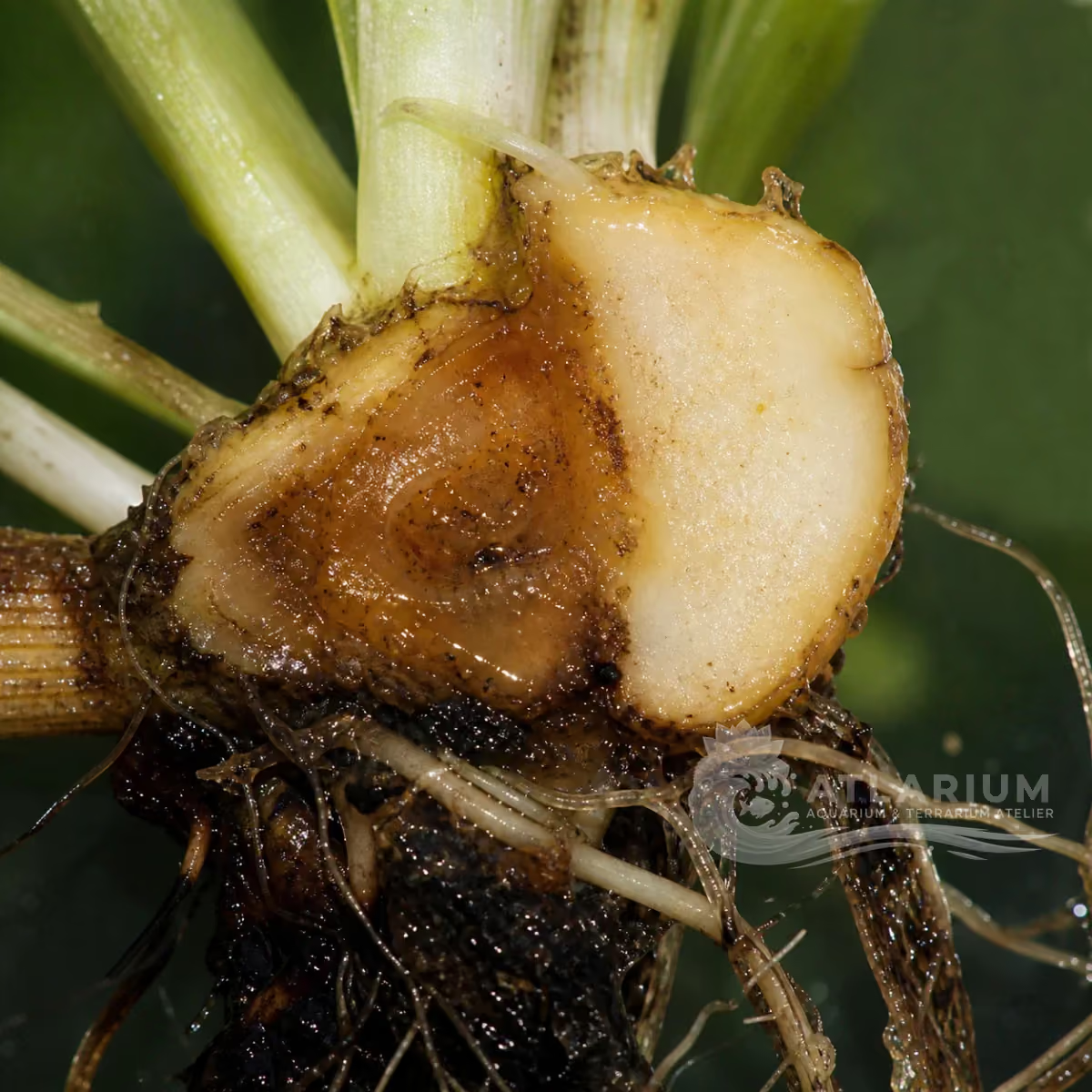
Motile aeromonads are an opportunistic bacterial complex associated with motile Aeromonas septicemia in fish. Reddening, ulcers, dropsy or mortality are compatible syndromes, not a species diagnosis. Pure culture and identification from infected tissue, ideally with susceptibility testing, are required before antimicrobial selection. Management begins by correcting stress, water quality, crowding and other predisposing factors while veterinary fish-health assessment directs any therapy.
Freshwater / Brackish
Gammaproteobacteria
Functional group
Low
Potential pathogen
Potentially harmful
Not cultured
No value supported by reliable evidence
Varies by taxon or life stage
No universal threshold supported by reliable evidence

Identity, habitat, and ecological role

Observation, culture, and ecological relationships

Geographic origin and distribution
Origin

Identification and observation data
- Record type
- Functional group
- This is a functional complex, not a single bacterial species. “Motile aeromonads” groups related opportunistic Aeromonas associated with motile aeromonad septicemia. The label is useful for a syndrome and laboratory workflow, but it cannot assign every ulcer, water bacterium or hemorrhage to one species. Species and strain identification require an appropriate clinical isolate.
- Recognition confidence
- Low
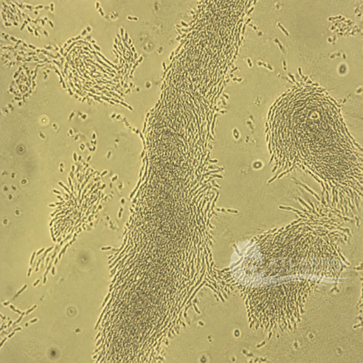
- Household microscopy may show small rods or aggregates but cannot identify Aeromonas, a motile complex or a resistance pattern. A defensible diagnosis starts with a pure culture from infected tissue, followed by identification and, when therapy is considered, antimicrobial susceptibility testing. Water samples or a photograph do not establish the cause of septicemia in an individual fish.
- Biological group
- Gammaproteobacteria
- Motile aeromonads are Gram-negative bacteria in Aeromonadaceae; the catalogue bacteria-and-archaea label is a broad navigation group, not a diagnostic method. Cells may be widespread in aquatic environments. The relevant distinction is between environmental presence, colonization and disease-causing isolates from affected tissue, rather than a visual bacterial category.
- Ecological functions
- Potential pathogen
- The documented role is potential opportunistic fish pathogen. Motile Aeromonas septicemia can involve hemorrhage, ulcers and systemic disease, but similar signs have several bacterial, parasitic, environmental and traumatic causes. Pathogenicity depends on strain and host context. The complex is not treated as a universal explanation for any red lesion or mortality event.
- Aquarium impact
- Potentially harmful
- A confirmed clinical isolate can be serious, especially with progressive ulcers, systemic signs or mortality. Its presence in water is not a diagnosis. Immediate priorities are fish-health assessment, containment, oxygenation and correction of stressors while diagnostic samples are obtained. Treating all fish with a guessed antibiotic can increase resistance and obscure the diagnosis.
- Culture and management
- Not cultured
- Motile aeromonads are not a home culture organism. Do not deliberately grow, move or exchange water, fish, lesions, filters or culture material from a suspect system. Use dedicated equipment, prevent cross-contamination and protect broken skin during handling. Culture and susceptibility testing belong to qualified diagnostic laboratories with appropriate biosafety and disposal.
- Typical size
- No value supported by reliable evidence
- The 0.3–4 μm guide describes bacterial-scale cells, not an identification criterion. Culture conditions, staining, preparation and the measured axis affect apparent size. A microscopy image may show cells or aggregates but cannot distinguish Aeromonas from many other bacteria. Record scale, sample type and method, then rely on culture, identification and susceptibility work for clinical claims.
- Visible to the naked eye
- Varies by taxon or life stage
- Individual bacterial cells are invisible to the naked eye. Cloudy water, slime, ulcers or hemorrhage are system or host signs and do not reveal which bacterium is involved. Only laboratory methods on appropriate clinical samples can identify a likely agent. Visible changes should trigger diagnosis and husbandry review rather than a name or drug choice.
- Useful starting magnification
- No universal threshold supported by reliable evidence
- Around 1000× with suitable staining may reveal bacterial morphology, but morphology cannot confirm Aeromonas, strain virulence or antibiotic susceptibility. Gram stain, culture, identification, PCR and susceptibility testing answer different questions. Use high magnification to document a sample, never to substitute for a clinical isolate when treatment, quarantine or mortality interpretation is at stake.

Links to Aquarium SOS


Microworld ecological network
Links based on documented kinship, food webs, biofilms, and ecological functions.