Freshwater velvet dinoflagellate
Piscinoodinium pillulare
Piscinoodinium pillulare is a freshwater parasitic dinoflagellate associated with velvet, rust or gold-dust disease in fish. Its pigmented trophonts attach to skin and gills, so the fine golden appearance is a useful warning but not a diagnosis by itself. Confirmation requires a wet mount of appropriate tissue, and treatment, containment and follow-up must account for the parasite’s off-host stages and for host sensitivity.
Water type
Freshwater
Biological group
Dinophyceae
Record type
Species
Recognition confidence
Medium
Ecological functions
Parasite, Potential pathogen
Aquarium impact
Potentially harmful
Culture and management
Not cultured
Typical size
30 µm – 150 µm
Visible to the naked eye
No, not as an individual
Useful starting magnification
400× to begin observation
Identity, habitat, and ecological role
Identity and taxonomic resolution: Piscinoodinium pillulare is a freshwater parasitic dinoflagellate associated with velvet, rust or gold-dust disease of fish. It must not be confused with the marine relative Amyloodinium merely because both cause similarly named syndromes. The old name Oodinium is used inconsistently in hobby material, so host salinity, sampling site and diagnostic characters are essential. A golden cast on a fish is a warning sign, not species proof or an automatic prescription.
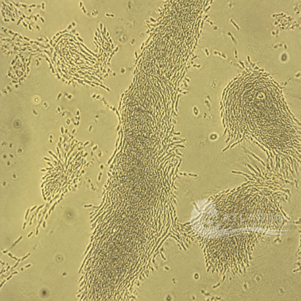
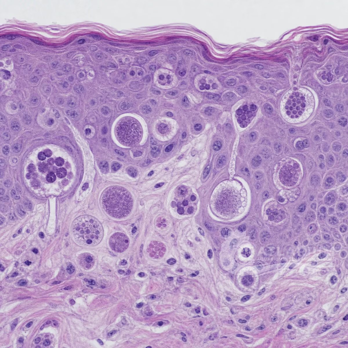
Appearance, attachment and dimensions: The pathogenic trophont is pigmented, nonflagellated and nonmotile while attached to skin or gills. Fine brown-golden dust, lethargy, reduced feeding, rubbing and respiratory distress can be compatible signs; gill-only infestations may lack obvious external spots. A wet mount of skin, fin or gill material can reveal the parasite and should be interpreted with host signs and other differentials. Note the 30–150 μm range as supportive morphology, not as a stand-alone identification.
Host sites and disease process: Attached trophonts invade or feed at the skin and gill surface. Heavy infestations can compromise gill function and can cause high mortality in cultured freshwater fish. Susceptibility and outcome vary by fish species, parasite burden, water quality and the delay before detection. Treat respiratory distress, rapid losses or diagnostic uncertainty as reasons for prompt aquatic-veterinary or laboratory assessment rather than waiting for color alone to settle the diagnosis.
Life cycle, spread and relevance: Mature attached stages form cysts that release small, flagellated free-swimming stages able to initiate new infections. A visibly improved fish therefore does not settle system risk. For a diagnostically supported case, the host, system and veterinary plan determine whether containment, movement limits or follow-up are appropriate; no universal quarantine or equipment rule follows. One suspicious fish alone does not prove this species caused every illness in the aquarium.
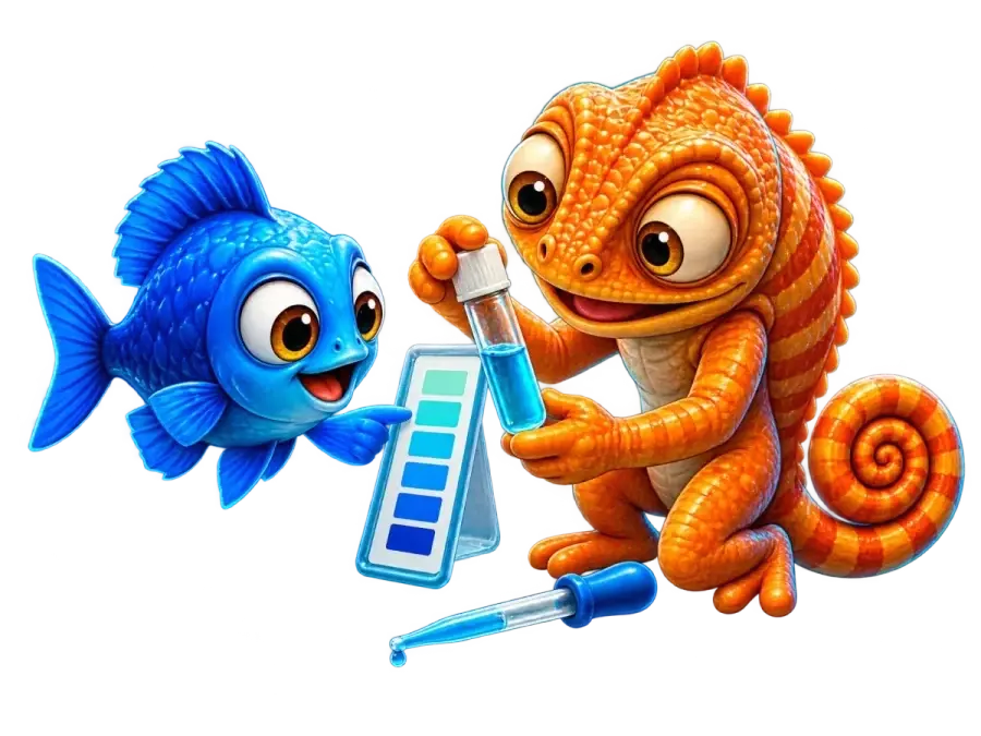
Observation, culture, and ecological relationships
Where to look and how to sample: Observe fish in good light for a fine golden or brown dust, rubbing, reduced feeding, lethargy, rapid breathing or surface piping, then sample the most relevant site. Skin, fin and especially gill wet mounts may be needed because infestation can be predominantly branchial. Note each slide with fish, body site, date and tank. Equipment handling is case-specific to the host, system and veterinary plan; one negative sample cannot exclude an early or focal infestation.
How to observe and identify: Start with a fresh wet mount and scan at low power before using about 400× to inspect suspected trophonts and the surrounding tissue. Compare several fields and fish: mucus, debris and other ectoparasites can mislead. Microscopy supports diagnosis only when it agrees with host signs, site and history. Severe gill disease, mortalities, atypical forms or uncertainty warrant veterinary or laboratory confirmation instead of assigning Piscinoodinium from a photograph.
Immediate containment and support: When compatible signs and diagnostic evidence identify a case, containment may be considered with the host, system and veterinary plan in view. Movement of fish, water, plants, nets or filter media, use of dedicated tools, handling and removal of dead fish need case-specific judgement rather than a blanket procedure. Check oxygenation and water quality, note the timeline, exposed species and signs, and keep any measures separate from diagnostic confirmation and treatment planning.
Treatment and professional limits: Veterinary references list copper sulfate for freshwater velvet, and a veterinarian may consider chloroquine for ornamental, non-food fish. Suitability and safety depend on host, water, formulation and authorization; any treatment product can injure fish or other aquarium organisms. No dose, duration or universal protocol is provided. Use only a legally labelled or veterinary-directed product after diagnosis, and follow its instructions and any food-fish restrictions exactly.
Control, follow-up and prevention: Cysts release infective free-swimming stages, so a cleaner fish alone does not settle risk. After veterinary treatment, a fish may need recheck about 7–10 days later; the clinician decides whether sampling, including a wet mount, suits that host and system. If recurrence is suspected, review diagnosis, source and husbandry context before considering quarantine, sanitation or movement measures. No universal duration, fallow interval, sanitation method or shared-equipment rule is established.
Risks, biosecurity and limits: Do not turn a gold sheen, a trade name or a web photograph into a diagnosis. Do not combine drugs, alter water chemistry or use a treatment product around sensitive species without qualified guidance. Protect staff by avoiding exposure to treatment chemicals, using appropriate gloves for broken skin and following product safety information. Syndrome-level conclusions remain conditional until clinical signs, microscopy and alternatives agree.
Geographic origin and distribution
Origin
North America
Identification and observation data
- Record type
- Species
- A species-level scientific name is used because the literature discusses Piscinoodinium pillulare specifically. That resolution applies only when diagnostic characters, host setting and freshwater context support it. A gold-colored film or an older trade name alone is insufficient, and a photograph of an aquarium organism can never establish the species. The published diagnostic sources do not establish one well-supported natural geographic origin for this species.
- Recognition confidence
- Medium
- Compatible signs can raise suspicion, but confirmation needs a wet mount of skin, fin or gill material and agreement with clinical history. Gill-heavy infestations can be visually inconspicuous, while mucus, debris and other ectoparasites can resemble the parasite. Laboratory or aquatic-veterinary help is appropriate for severe disease, mortality or unclear findings.
- Biological group
- Dinophyceae
- Piscinoodinium is a dinoflagellate, not a ciliate or a generic “alga” diagnosis. Its attached pathogenic trophonts are pigmented, nonmotile and nonflagellated; later cyst stages release small flagellated infective stages. The taxonomic context provides biological context, but it does not erase the need to distinguish freshwater Piscinoodinium from the marine velvet agent Amyloodinium. The published freshwater diagnostic sources confirm the taxonomic context but do not establish a well-supported order for this species.
- Ecological functions
- Parasite, Potential pathogen
- The documented ecological roles are parasite and pathogen because trophonts attach to the skin and gills of compatible freshwater fish and can damage those tissues. Clinical significance depends on burden, host susceptibility, water quality and stage. An environmental finding, a symptom or a positive sample from one fish does not automatically establish that this organism is the sole cause of illness throughout a tank.
- Aquarium impact
- Potentially harmful
- This organism is potentially harmful because heavy infestations can impair gill function and cause substantial mortality. A case with respiratory distress or losses needs diagnostic sampling and professional assessment. For a diagnostically supported case, the host, system and veterinary plan determine whether any containment or intervention is appropriate. Color alone is not a basis for medication, and correct identification, water chemistry, cohabitants and regulation affect safety.
- Culture and management
- Not cultured
- No domestic culture or propagation method is provided because the cited evidence addresses fish disease rather than maintenance of this organism. An accidental observation does not identify a culture method or establish that material can be safely handled. For a diagnostically supported case, the host, system and veterinary plan determine whether sampling, isolation or other handling is appropriate; no universal containment procedure follows.
- Typical size
- 30 µm – 150 µm
- A reported working size of 30–150 μm can help frame microscopic observations, but trophont size varies with stage, host site and preparation. Measure intact material with a scale, state what structure was measured and preserve the image context. The range supports a differential; it cannot distinguish Piscinoodinium from every similarly sized particle, cell or ectoparasite by itself.
- Visible to the naked eye
- No, not as an individual
- Individual parasite stages are not reliably identifiable with the naked eye. A fine golden or brown dust on a fish may become visible, but that appearance is a clinical clue rather than a direct view of a named organism. Gill-localized infestation can produce respiratory signs without conspicuous spots, so absence of a visible coating cannot safely rule out the problem.
- Useful starting magnification
- 400× to begin observation
- Begin a fresh wet mount at low power to locate relevant material, then use about 400× to inspect trophonts and the surrounding tissue. Examine several fields and, when possible, more than one affected fish. Magnification alone does not provide diagnosis: site, morphology, host signs and alternative causes must be reconciled, with laboratory or veterinary confirmation for consequential decisions.
Links to Aquarium SOS

Microworld ecological network
Links based on documented kinship, food webs, biofilms, and ecological functions.