Cryptocaryon irritans
Cryptocaryon irritans
Cryptocaryon irritans is a marine ciliate associated with white spot disease only when confirmed in compatible fish tissue. Visible white spots can overlap with other causes, and gill-only infection may show no spots. Its trophont, protomont, tomont and theront stages make management stage dependent. Use skin, fin and gill microscopy with fish and system context; treatment, control and prevention require qualified advice, not a universal drug, dose, duration or fallow interval.
Water type
Marine
Biological group
Prostomatea
Record type
Species
Recognition confidence
Medium
Ecological functions
Parasite, Potential pathogen
Aquarium impact
Potentially harmful
Culture and management
Not cultured
Typical size
100 µm – 500 µm
Visible to the naked eye
Yes, as an individual
Useful starting magnification
100× to begin observation
Identity, habitat, and ecological role
Identity and taxonomic resolution: Cryptocaryon irritans is a marine ciliate identified at species level only from compatible fish tissue, not from every white spot. UF/IFAS describes marine white spot disease in wild and cultured marine fishes, but similar marks, mucus and respiratory signs have other causes. Gill-only infection may have no visible spots. Confirmation joins the fish, sampled skin, fin or gill tissue, a wet mount and compatible stage; retain a differential when those observations are incomplete.
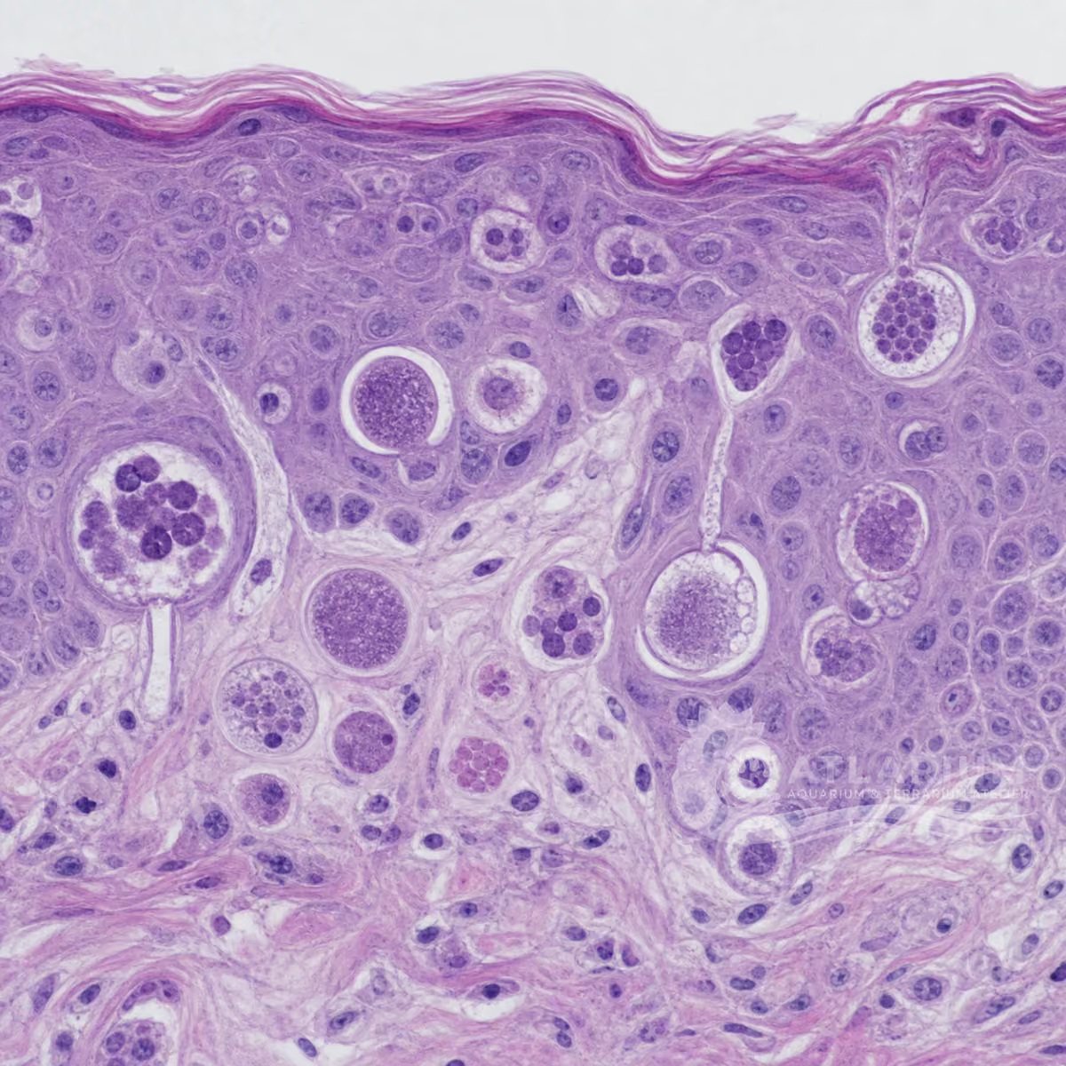
Morphology and dimensions: The observed 100–500 μm working range is a stage-dependent guide, not a diagnostic boundary. Trophonts embedded in skin or gill epithelium can be spherical, club- or pear-shaped, ciliated and slowly rolling; their opaque cytoplasm can hide the lobed macronucleus. Protomonts, encysted tomonts and free-swimming theronts differ in form and exposure. Preserve sample site, scale, preparation and movement because size, strain, host, salinity and temperature affect interpretation.
Habitat and microhabitat: This marine parasite has a direct lifecycle across fish tissue and the surrounding system. Trophonts feed in the host; after leaving, protomonts settle and encyst as tomonts on surfaces; tomites divide and emerge as theronts that seek fish. Water, substrate, tank structures, equipment and host tissue therefore differ in their roles in observation and transfer. A parasite seen at one site does not demonstrate infection throughout a system or identify a geographic origin.
Food-web role and aquarium meaning: C. irritans is an obligate fish parasite and potential pathogen, not a cleanup organism or live food. Disease severity can vary with parasite strain, fish species and condition, temperature, salinity, oxygen and exposure. A visible spot, dead fish or environmental stage does not prove a tank-wide burden or causal diagnosis. Keep compatible signs, confirmed parasite, host stressors and alternative diagnoses distinct when judging urgency, containment or the risk to other fish.

Observation, culture, and ecological relationships
Where and how to sample: Collect fresh skin, fin and gill material from fish with compatible signs, keeping sites separate and noting fish, date, system, recent additions, salinity, temperature, oxygen and measured water conditions. A skin scrape, fin sample, gill biopsy and water drop recover different material. Use clean dedicated tools and do not transfer water or mucus between aquariums.
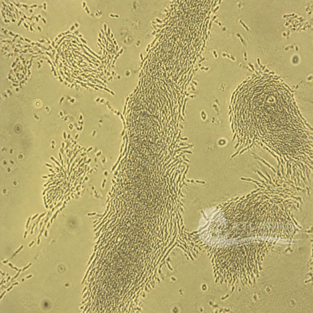
How to observe and identify: Begin with a fresh wet mount at low power, then increase magnification to examine ciliation, shape, movement and stage. Trophonts may roll under epithelium, while free theronts and other ciliates can overlap in appearance. Compare several fields and skin, fin and gill material when warranted. White spots or a photograph do not establish Cryptocaryon. Fish-health support or laboratory confirmation is appropriate when identity changes quarantine, movement, welfare or treatment decisions.
Reading a finding in the aquarium: Interpret microscopy with fish behavior, respiratory effort, lesion distribution, abundance, trend and measured water quality. Gill involvement can be serious even without external spots, while the same visible sign can have non-Cryptocaryon causes. Stabilise safe husbandry, support oxygenation and document changes while the differential is assessed.
Culture, containment and transfer: C. irritans is not a home culture organism and no propagation protocol is provided. Do not maintain, transfer, aerosolise or deliberately multiply suspect fish, water, substrate or material. Restrict affected-fish movement when appropriate, use dedicated nets and tools, and prevent shared water or mucus transfer. Quarantine, diagnostic isolation and experimental maintenance belong to qualified fish-health settings, where biosecurity and animal condition can be evaluated together.
Treatment, control and prevention: Treatment is not a universal product, dose, duration or fallow interval. Trophonts in tissue and protected tomonts are not equivalent to free-swimming theronts, the more exposed stage. UF/IFAS discusses copper, chloroquine, salinity change and formalin as context-dependent options with monitoring, host tolerance, invertebrate toxicity, legal and system limits. Confirm diagnosis and lifecycle timing, then seek qualified fish-health direction.
Limits, documentation and specialist referral: Keep fish identity, tissue site, signs, dates, water values, sample method, images, microscopy and actions together in the case documentation. Do not prescribe medication, certify an aquarium free of infection or apply one regimen to reef, mixed, food-fish and display systems. Escalate progressive disease, respiratory compromise, mortality, uncertain identification or movement decisions to an aquatic veterinarian or qualified laboratory.

Geographic origin and distribution
Origin
Tropical oceans and reefs
Identification and observation data
- Record type
- Species
- Cryptocaryon irritans is the marine ciliate associated with white spot disease. A white mark does not establish the species: other parasites, injury and noninfectious changes can overlap, while gill infection may not create visible spots. Confirm by wet mount of suitable tissue and retain host, sample site and life-stage context. The scientific name is not a substitute for a diagnostic sample. No geographic origin is shown because a marine disease context does not establish a well-supported geographic origin.
- Recognition confidence
- Medium
- Confidence is moderate only after wet-mount microscopy of skin, fin or gill tissue shows compatible parasites. White spots, flashing, respiratory difficulty and mortality are compatible observations, not species characters. Examine several sites because parasite distribution and stage vary. A single image cannot distinguish Cryptocaryon from other causes. Seek fish-health support if the result determines treatment, quarantine, mortality interpretation or movement of marine fish.
- Biological group
- Prostomatea
- Cryptocaryon irritans belongs to the marine ciliates in class Prostomatea, within the broader aquarium context of protists and infusoria. This relationship explains why microscopy and life-stage interpretation matter, but not all ciliates share the same lifecycle, host impact or treatment response. Compare morphology in a wet mount; a white spot alone is not evidence of this species. The available sources support the species, genus, family and class but do not establish an order.
- Ecological functions
- Parasite, Potential pathogen
- Parasite and pathogen are supported roles for confirmed Cryptocaryon irritans on compatible marine fish. Trophonts feed in skin and gills, while stages leaving the host develop on substrate before infective stages emerge. The clinical outcome depends on burden, host, water conditions and stage. Do not infer a tank-wide burden or causal role from one spot, a dead fish or water alone; connect findings through wet mount and time-course evidence.
- Aquarium impact
- Potentially harmful
- Impact is potentially harmful because confirmed infection can damage skin and gills, compromise respiration and multiply rapidly through the lifecycle. A suspect fish warrants prompt assessment, but visual spots do not establish an emergency treatment protocol for every resident. Isolate as appropriate, support oxygenation and monitor water quality. Copper can harm invertebrates, plants and biofilters, so a community or reef tank must not be medicated on a presumed species.
- Culture and management
- Not cultured
- This parasite is not cultured at home. Diagnostics should use fresh affected tissue, appropriate microscopy and containment under fish-health guidance. Do not transfer suspect fish, water, nets, substrate or equipment to another aquarium. Management focuses on quarantine, dedicated equipment, observation and safe care of affected animals. A laboratory wet mount is not permission to maintain the parasite or reuse a sample as a culture.
- Typical size
- 100 µm – 500 µm
- The 100–500 µm range is a broad guide across observable Cryptocaryon stages, not a stand-alone diagnostic boundary. Parasite size changes with stage, preparation and measurement method, and other protists can overlap. Measure a suitable wet-mount organism with scale and document the tissue site. Size alone cannot identify Cryptocaryon, establish lifecycle stage or determine the duration or safety of treatment.
- Visible to the naked eye
- Yes, as an individual
- Some host-associated cysts may be visible as white spots, but individual identity and gill involvement require magnification. A fish can have gill infection without obvious external spots, and visible spots may have other causes. Document the number, location and time course without scraping aggressively. A spot’s visibility does not certify Cryptocaryon, show all stages or identify a safe medication for the fish and system.
- Useful starting magnification
- 100× to begin observation
- About 100× total magnification is a useful starting point for locating larger compatible parasites in a skin, fin or gill wet mount; higher magnification and expert comparison may be needed for confirmation. Document site, preparation, scale and signs because stage matters. The magnification is an observation guide, not a treatment threshold. Veterinary direction should connect confirmed identity, lifecycle, fish sensitivity and system constraints before intervention.
Links to Aquarium SOS

Microworld ecological network
Links based on documented kinship, food webs, biofilms, and ecological functions.