Amphibian chytrid fungus
Batrachochytrium dendrobatidis
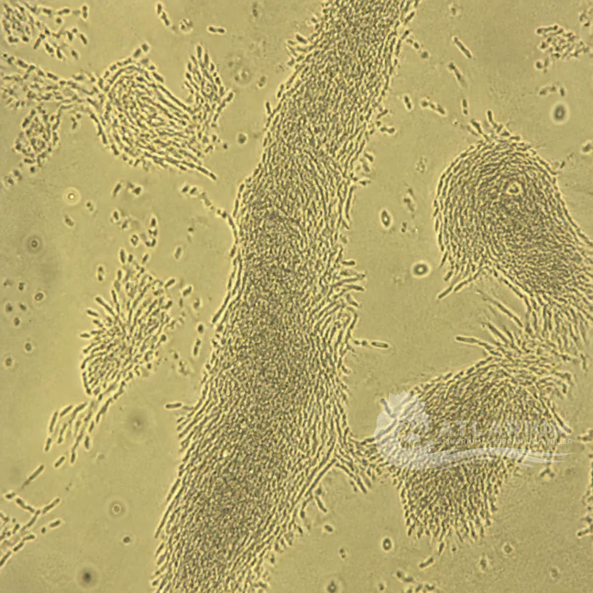
Batrachochytrium dendrobatidis is the zoosporic chytrid fungus associated with chytridiomycosis in amphibians. It is a high-biosecurity pathogen that may be reportable depending on jurisdiction: visual signs or a water observation are not a diagnosis. Confirm compatible cases through validated laboratory methods, isolate animals and equipment, and involve an amphibian veterinarian, laboratory and applicable authority. Treatment can be species-specific veterinary care; no dose, home protocol or culture method is provided.
Freshwater / Semi-aquatic
Chytridiomycetes
Species
Medium
Parasite, Potential pathogen
Potentially harmful
Not cultured
3 µm – 40 µm
No, not as an individual
400× to begin observation
Identity, habitat, and ecological role
Observation, culture, and ecological relationships
Identification and observation data
- Record type
- Species
- Batrachochytrium dendrobatidis is the zoosporic chytrid fungus associated with amphibian chytridiomycosis. Skin change, shedding or mortality do not identify the fungus. Use the name only when susceptible host, appropriate sample and validated laboratory evidence agree. A photograph, syndrome name or assumed exposure cannot confirm infection or replace reporting and veterinary decisions. No single geographic origin can be assigned because the reviewed institutional and veterinary sources do not establish one well-supported origin for this pathogen.
- Recognition confidence
- Medium
- Clinical signs and skin changes can raise suspicion, but Bd cannot be assigned from appearance alone. Validated sampling, histology or molecular methods are used for confirmation, and other amphibian diseases remain differentials. Confidence remains moderate so that an observation is not mistaken for certainty about species, causation or treatment. In a high-consequence setting, consult a qualified amphibian veterinarian or laboratory before moving animals or material.
- Biological group
- Chytridiomycetes
- Bd is a chytrid fungus in the Chytridiomycetes; oomycetes belong to a distinct lineage. Its free-swimming zoospores and skin-associated stages are not visible signs that identify the species in an enclosure. Freshwater and semi-aquatic microhabitats, host skin and equipment are distinct contexts. Microscopy can support suspicion; validated laboratory methods establish the agent in a health investigation.
- Ecological functions
- Parasite, Potential pathogen
- The documented roles are parasite and pathogen because Bd infects keratinized amphibian tissues and can cause potentially fatal chytridiomycosis. Susceptibility, life stage, host species, burden and environmental context affect disease expression. Detection or exposure does not prove clinical disease in every individual, while a compatible syndrome does not prove Bd. Keep host-level evidence, diagnosis and management decisions separate.
- Aquarium impact
- Potentially harmful
- The potential impact is harmful because Bd can spread among susceptible amphibians and has serious conservation and captive-population consequences. It is not a reason for unverified testing, home culture or mass treatment. Isolate suspect animals and equipment, document movements and seek veterinary, laboratory and competent-authority guidance. A diagnosis must guide the response; visual similarity never establishes a reportable infection.
- Culture and management
- Not cultured
- Bd is not a home culture. Isolation, maintenance and diagnostic testing belong to qualified laboratories with appropriate containment and regulatory awareness. Do not grow, enrich, transfer or share samples or materials of unknown origin from skin, water, substrate or equipment between amphibian systems. Dedicated tools, quarantine and controlled disposal limit spread while an investigation proceeds. Survival in water or on a surface does not make material safe to retain or use.
- Typical size
- 3 µm – 40 µm
- Relevant fungal stages fall within a 3–40 μm working range, but size varies with life stage and preparation. The range provides microscopic scale, not an infection or species diagnosis. Document sample type, host site and method rather than measuring an environmental particle in isolation. Dimensions cannot determine viability, burden, host susceptibility or the need for a treatment decision.
- Visible to the naked eye
- No, not as an individual
- Individual fungal stages are not reliably identifiable to the naked eye. Skin shedding, dull colour, lethargy or abnormal posture may be visible in an amphibian but are not a Bd label and have other causes. An enclosure surface, water change or residue does not demonstrate contamination. Use clinical context and validated testing; appearance alone cannot establish disease, transmission risk or clearance.
- Useful starting magnification
- 400× to begin observation
- At around 400×, the observer can examine relevant structures in prepared material, but microscopy alone cannot provide a definitive Bd result in a health investigation. Preserve the host site, sample method and chain of information. Confirmation uses validated laboratory approaches such as those in the WOAH manual. Magnification cannot establish the safe movement of animals, the correct treatment or the absence of infection.
Links to Aquarium SOS

Microworld ecological network
Links based on documented kinship, food webs, biofilms, and ecological functions.