Marine velvet dinoflagellate
Amyloodinium ocellatum
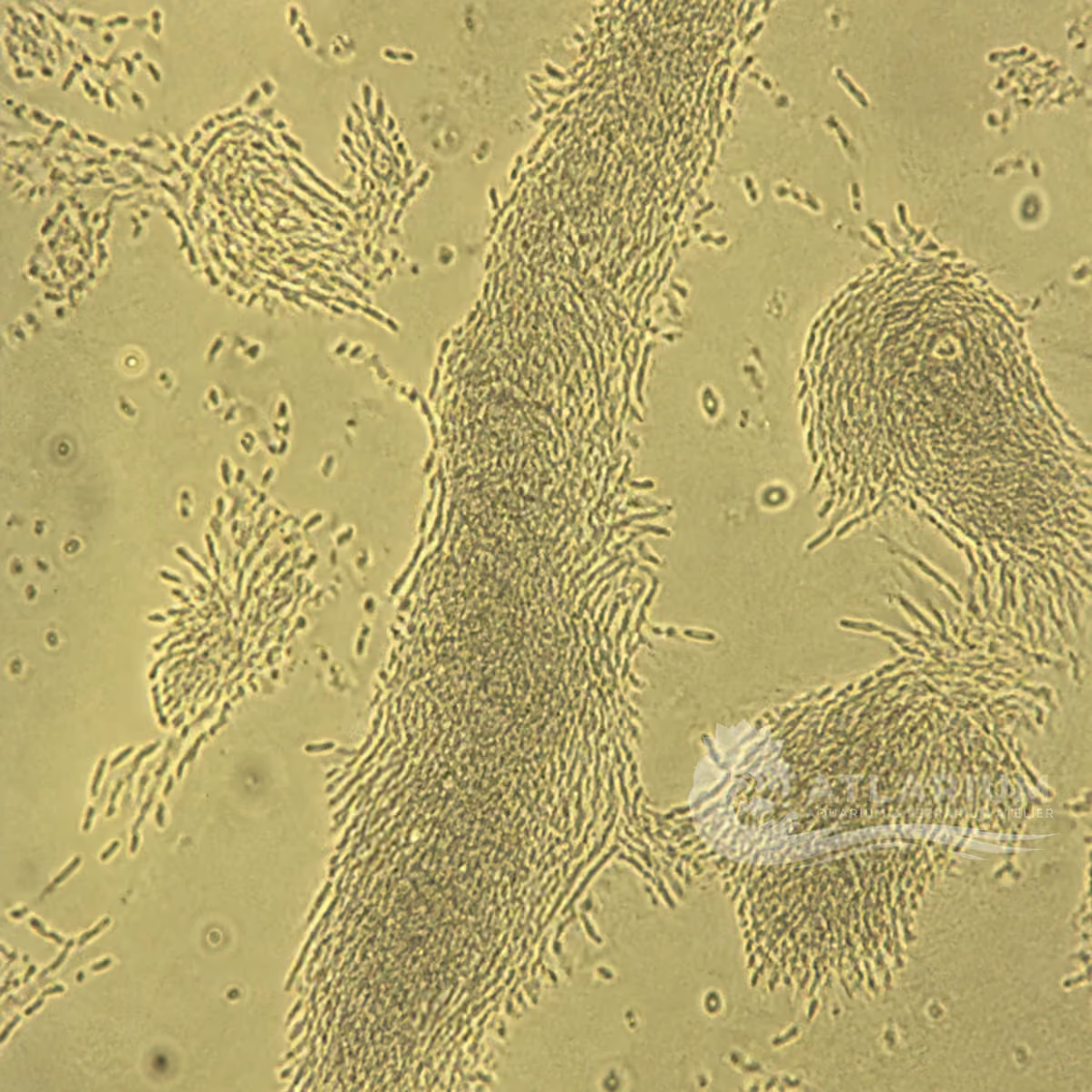
Amyloodinium ocellatum is a parasitic dinoflagellate of marine and brackish-water fish. Its direct cycle includes an attached feeding trophont, an encysted tomont and a free-swimming infective dinospore. A wet-mount finding can support diagnosis in affected fish with compatible clinical signs, but a visible velvet-like appearance or a photograph alone does not establish cause or treatment.
Water type
Brackish / Marine
Biological group
Dinophyceae
Record type
Species
Recognition confidence
Medium
Ecological functions
Parasite, Potential pathogen
Aquarium impact
Potentially harmful
Culture and management
Not cultured
Typical size
13 µm – 350 µm
Visible to the naked eye
No, not as an individual
Useful starting magnification
40× to begin observation
Identity, habitat, and ecological role
Identity and nomenclature: Amyloodinium ocellatum is a species of dinoflagellate placed by the NCBI taxonomy lineage in Dinophyceae, Thoracosphaerales and Thoracosphaeraceae. Oodinium ocellatum is its basionym, so older reports may use that name. The taxon should not be confused with freshwater Piscinoodinium or with other organisms that produce spots, haze or excess mucus. Species-level identification requires compatible host tissue and diagnostic observation; resemblance in aquarium water alone is not enough.
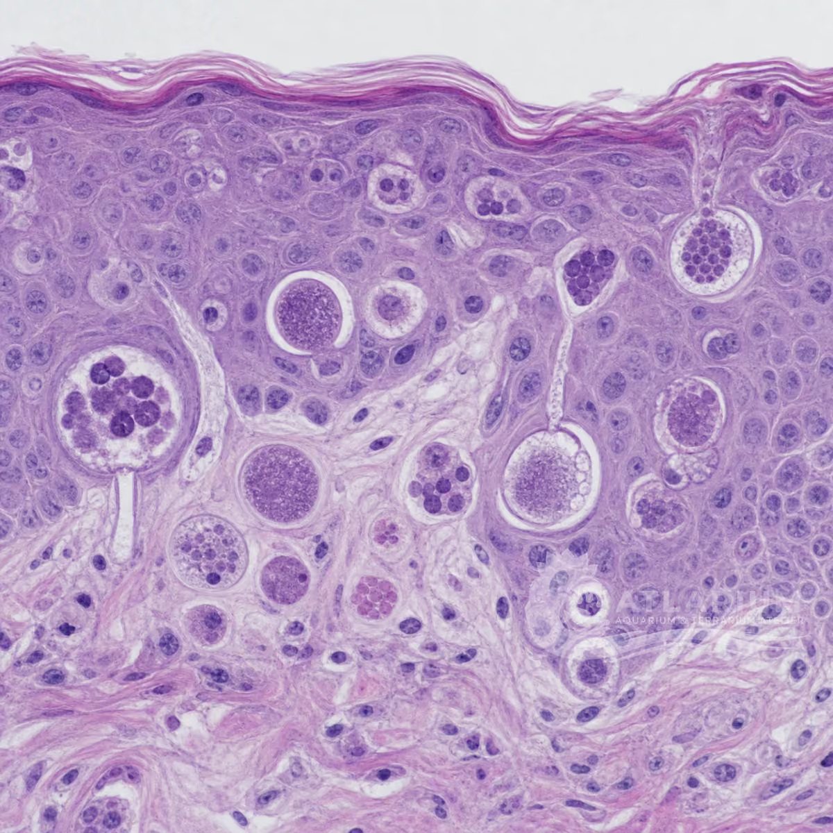
Life cycle and form: The direct cycle has three documented functional stages. The trophont feeds while attached to fish epithelium by rhizoids; after leaving the host it becomes an encysted, dividing tomont on a substrate; dinospores released from the tomont swim to find another host. Cycle duration, trophont size and tomont output vary with temperature and other conditions. University sources report mature trophonts averaging about 80–100+ µm and a tomont can form up to 256 infective stages.
Host and environmental context: This parasite is associated with marine and brackish-water fish. Attached stages can occur on gills, skin, fins and body surfaces, while the encysted stage settles onto a tank surface or substrate. Gill-only infections may lack the externally visible velvet appearance. The available evidence supports a direct parasite–fish cycle, not a harmless free-living protist role, so an observation should be interpreted with the host, affected tissue and recent system history.
Meaning of a finding: Amyloodiniosis, often called marine velvet disease, can be serious and may progress rapidly in affected fish. Lethargy, poor feeding, rubbing, altered respiration or a brownish-gold haze are compatible signs, but none is unique to this taxon. Wet mounts of affected tissue provide useful evidence, and molecular or veterinary confirmation can resolve uncertainty. Document the host, tissue, salinity and water values; do not infer a diagnosis, contagion pathway or medicine from one visual sign.
Observation, culture, and ecological relationships
Where and how to observe: Observation is meaningful when it starts at the affected fish and tissue, not from a nonspecific water sample. A wet mount of gill, skin or fin material may show attached stages, whereas a water-only sample selects different life stages. Preserve the context with date, fish species, tissue, salinity and temperature. Samples taken from a marine or brackish fish should be handled in water of comparable salinity for examination, because an abrupt freshwater exposure can detach the parasite.
Microscopy and discrimination: For tissue wet mounts, university guidance reports that attached trophonts can be visible at 40× or 100× total light-microscope magnification; 400× can add detail but is not the minimum for finding them. Trophonts may be dark brown, ovoid to pear-shaped and non-motile while attached. Compare more than one field and distinguish them from Cryptocaryon irritans. Retain the highest taxonomic resolution actually supported by the preparation and avoid naming a species from a distant photograph.
Interpreting aquarium risk: An affected host and compatible wet-mount finding merit timely assessment because gill involvement can compromise respiration. A visible golden dust effect is not required when infection is confined to gills, and its presence is not proof by itself. Treat observations as a fish-health concern: isolate the interpretation from assumptions about water quality, then obtain qualified veterinary or laboratory advice for illness, spread or mortality.
Treatment and stage limits: University and veterinary references report chemical treatment as one control option: formalin or hydrogen peroxide for cited food-fish contexts, and copper sulfate or chloroquine for ornamental fish. The attached trophont and encysted tomont are not equally susceptible, so treatment must be planned against the cycle rather than presumed to clear every stage at once. Product label, host species, food-fish status and professional monitoring determine whether an option is appropriate.
System control and prevention: Control is not limited to medicine. For aquaculture systems, the documented approaches include flushing to remove bottom-settled tomonts, filtration to remove that stage, and ultraviolet irradiation or ozone to reduce parasite numbers. Prevention also relies on quarantine of new fish, isolation of affected populations, dedicated equipment and sanitation. These measures reduce transmission opportunity but do not replace examination of fish or verify that an individual system is free of infection.
Limits, documentation and specialist referral: Useful case documentation identifies the fish, system, sample site, observed signs, water conditions, images and microscopy method. Chemical choice, concentration, duration, repeat schedule and discharge rules are not universal: copper can harm invertebrates and biofilters, and some options are not permitted for food fish. Share the original observation with an aquatic veterinarian or diagnostic laboratory; progressive respiratory signs, spread or mortality need prompt case-specific assessment.
Geographic origin and distribution
Origin
North America
Identification and observation data
- Record type
- Species
- Amyloodinium ocellatum is a named species, rather than an informal aquarium appearance. Its older basionym is Oodinium ocellatum, so historical sources can use a different combination. A finding on fish must still be supported by compatible tissue, morphology and context; a photograph of haze, spots or water particles does not establish this species. No single geographic origin can be assigned because the reviewed taxonomy and fish-health sources do not establish one well-supported origin for this species.
- Recognition confidence
- Medium
- The confidence is deliberately moderate because affected tissue can support a preliminary microscopic identification, but look-alikes and preparation quality matter. Attached dark trophonts in a suitable wet mount are more informative than a distant image. Where disease management depends on the answer, an aquatic veterinarian or diagnostic laboratory should decide whether additional microscopy, histology or molecular testing is needed.
- Biological group
- Dinophyceae
- Amyloodinium ocellatum is a dinoflagellate. Current NCBI taxonomy places it in the class Dinophyceae, order Thoracosphaerales and family Thoracosphaeraceae. The broad aquarium term “protist” is useful for discovery but is not a diagnosis and should not be used to transfer the ecology of unrelated ciliates, amoebae or free-living dinoflagellates to this fish parasite.
- Ecological functions
- Parasite, Potential pathogen
- The documented roles are parasite and pathogen in compatible marine and brackish-water fish. The feeding trophont attaches to host epithelium by rhizoids, while the tomont and dinospore are non-attached stages in the direct cycle. It should not be described as a normal biofilm grazer, cleanup organism or harmless planktonic companion merely because some other dinoflagellates are free-living.
- Aquarium impact
- Potentially harmful
- Its aquarium impact is potentially harmful because compatible infections can damage gill and skin tissue and may lead to serious disease or mortality. The impact depends on a real host-associated infection, not simply on a suspected visual resemblance in the water. Gill-only infection may lack an obvious velvet coating, so absence of visible dust does not exclude concern when compatible respiratory signs are present.
- Culture and management
- Not cultured
- This species is not offered as a home culture. Its direct cycle requires fish-associated stages, and propagating or transferring uncertain material would create a biosecurity risk rather than a useful observation exercise. Diagnostic isolation, confirmation and any controlled maintenance belong to qualified facilities. For aquariums, the appropriate aim is careful observation, containment and professional assessment rather than enrichment or multiplication.
- Typical size
- 13 µm – 350 µm
- Size depends on life stage and conditions. A 2025 study measured sampled dinospores at 13.03–19.66 µm long, while university extension material reports trophonts commonly below 150 µm and possible maximum trophont sizes of 350 µm. The 13–350 µm range therefore spans documented stages; it is not the expected size of every organism in one wet mount.
- Visible to the naked eye
- No, not as an individual
- Individual stages are normally assessed with microscopy. A fish may show a brownish-gold, dusty or velvety appearance when skin is involved, but that visible effect is not the individual parasite and is not species confirmation. If infection is confined to gills, the external effect may be absent. Observation must therefore combine host signs and suitable tissue examination rather than rely on naked-eye appearance.
- Useful starting magnification
- 40× to begin observation
- For gill or skin wet mounts, university extension guidance reports that attached trophonts can be seen at 40× and 100× total light-microscope magnification. Higher magnification, including 400×, may help inspect detail but is not required as the first detection threshold. A magnification value describes observation conditions, not proof of species identity; comparable tissue and differential assessment remain essential.
Links to Aquarium SOS

Microworld ecological network
Links based on documented kinship, food webs, biofilms, and ecological functions.