Chilodonella spp.
Chilodonella
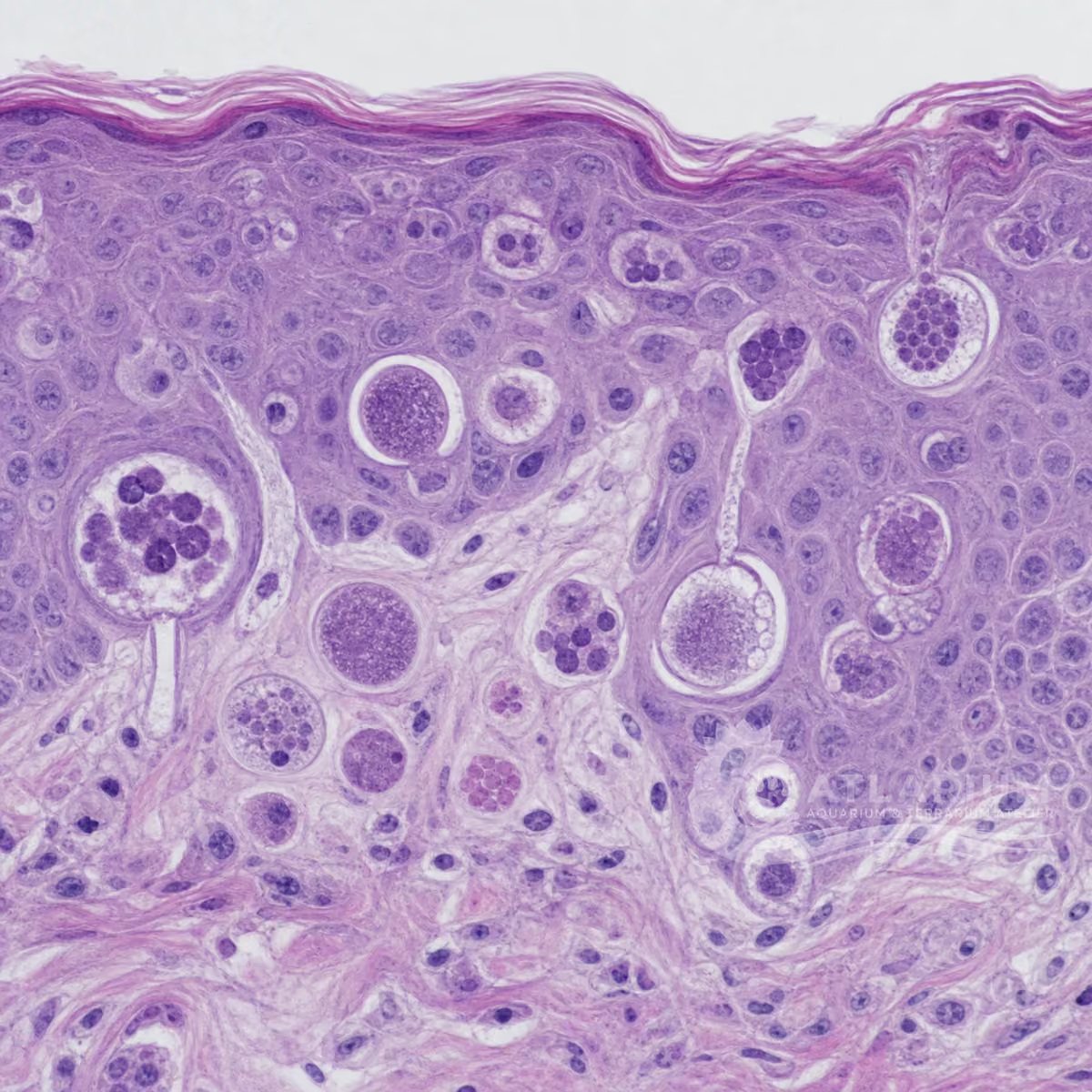
Chilodonella is a freshwater-context genus of ciliates; compatible fish-associated infestation may involve skin, fins or gills. Excess mucus, flashing, loss of condition and respiratory signs are reasons to assess affected tissue, not a species diagnosis. The 30–80 µm range is only a wet-mount guide. Treatment belongs after confirmation, fish-specific safety and applicable veterinary and regulatory review, never from a visible sign alone.
Water type
Freshwater
Biological group
Phyllopharyngea
Record type
Genus
Recognition confidence
Low
Ecological functions
Parasite, Potential pathogen
Aquarium impact
Potentially harmful
Culture and management
Not cultured
Typical size
30 µm – 80 µm
Visible to the naked eye
No, not as an individual
Useful starting magnification
No universal threshold supported by reliable evidence

Identity, habitat, and ecological role
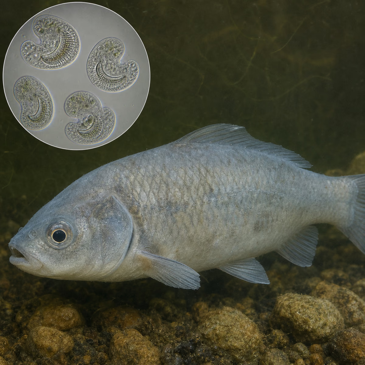
Identity and taxonomic resolution: Chilodonella is treated as a genus in Chilodonellidae and Phyllopharyngea under the catalog convention. The card does not turn every flattened ciliate, mucus film or fish symptom into a Chilodonella species diagnosis. Ovoid or kidney-like form, dorsoventral flattening, cilia and movement can orient a wet-mount comparison, but related organisms and preparation effects remain part of the differential. Retain host, tissue and sample context when a finding needs interpretation.
Morphology and dimensions: UF/IFAS gives an approximately 30–80 µm by 20–60 µm visual guide for Chilodonella in fresh fish samples. The stored 30–80 µm range therefore helps scale a wet mount, not identify a species, quantify burden or choose treatment. Cell posture, preparation, tissue and overlapping small ciliates affect what is seen. Record several intact organisms with a calibrated scale and the affected site rather than relying on one image, size or compressed specimen.
Freshwater and host microhabitat: Freshwater is the fish-health context of this card, not a global distribution, genus-origin or salinity-transfer claim. Compatible fish-associated findings may concern skin, fins and gills, which are different sample sites from water or detritus. A fresh tissue sample can contain mucus, host cells and other organisms as well as a ciliate. Do not extrapolate one wet mount to the whole system or use it to infer a generic aquatic habitat rule.
Host relationship and clinical meaning: Parasite and pathogen labels apply here only to compatible fish-associated infestation. Excess mucus, flashing, poor condition, altered gills and respiratory distress can occur, but they are host signs rather than species-level evidence or proof of causation. Microscopic examination of affected tissue or a wet mount supplies diagnostic context; it does not eliminate differential diagnoses by itself. An environmental observation cannot establish infestation, clinical load or a treatment need.

Observation, culture, and ecological relationships
Where to look and how to sample: Treat mucus, flashing or respiratory change as a reason to obtain a fresh assessment of affected tissue, not as a name for the agent. Record the fish, sample site, timing, water context and handling history, then preserve skin, fin and gill material separately. A wet mount can support observation of motile ciliates, host cells and debris in that sampled material. It cannot establish an absence elsewhere, identify every organism present or convert a sign on one fish into a system-wide diagnosis.
How to observe and identify: Examine fresh affected material with a calibrated scale and compare several intact organisms, fields and tissue sites. A flattened, free-moving, ovoid or kidney-like ciliate can support a cautious Chilodonella orientation, but more evidence may be needed. No fixed magnification value is stored: 400× may aid observation but is not a sufficient universal diagnosis threshold. Do not infer identity, burden or treatment eligibility from one photograph, one size or mucus alone.
Presence and meaning in an aquarium: A compatible ciliate finding should be read with the fish, tissue, signs and water conditions. It is not automatically the first cause of poor condition, a verdict on water quality or proof that every fish is infested. Freshwater describes the supported clinical context, not a full range or salinity instruction. Visible mucus, dull colour, altered gills and rapid breathing remain non-diagnostic until fresh-tissue microscopy and qualified interpretation correlate them.
Culture, maintenance or use: Chilodonella is not offered as a home or feeder culture. Keep suspect material out of transfers and do not attempt propagation. The evidence supports retaining sample and fish context, not an experimental culture recipe, survival target or decontamination schedule. If diagnostic handling is needed, use qualified fish-health or laboratory procedures that match the affected host and facility. A container of water or a mucus film does not establish a stable or safe culture.
Documented ecological relationships: The supported relationship is host-associated and conditional: compatible freshwater fish infestation can involve skin, fins and gills. It does not show every genus member or mucus response is pathogenic. Species, tissue burden, host condition and co-infections can change clinical meaning. Keep parasite and pathogen labels tied to confirmed compatible infestation rather than transferring them to free water, debris or an unexamined host.
Risks, control and limits: Do not self-prescribe medication, copper, formalin, salt, baths, temperature change or a fixed quarantine schedule from a sign or image. Merck's categories follow diagnosis; this card supplies no product or dose. While awaiting a fresh affected-tissue assessment, contain suspect fish and avoid moving fish, water, nets or sampling equipment. Treatment requires confirmation plus host safety, formulation and veterinary/regulatory review; supportive care does not identify or clear the ciliate.

Geographic origin and distribution
Origin
Cosmopolitan or introduced

Identification and observation data
- Record type
- Genus
- This is a genus-level entry for Chilodonella, not a diagnosis of one species or every flattened ciliate on a fish. The stored classification follows Chilodonellidae and Phyllopharyngea; no order scalar is chosen because rank treatment is authority-dependent. A wet-mount appearance can orient comparison, but host, tissue, several intact organisms and diagnostic evidence determine the supported resolution. Keep a finding tied to its fish, sample site and clinical context.
- Recognition confidence
- Low
- Identification confidence is medium only for a cautious genus orientation from fresh affected tissue, multiple intact organisms, a scale and compatible morphology. Ovoid or kidney-like, dorsoventrally flattened, motile cells can suggest Chilodonella, but a photograph, mucus response or one field cannot resolve species or causation. No fixed magnification is diagnostic. Use suitable specialist evidence when a result changes fish movement, health interpretation or treatment decisions.
- Biological group
- Phyllopharyngea
- Chilodonella is handled in the protists-and-infusoria navigation group and is a ciliate genus in Chilodonellidae and Phyllopharyngea under the catalog convention. The group label helps distinguish a microscopic ciliate from the host's mucus response, not identify all ciliates as the same organism or disease risk. Use it to guide a cautious wet-mount differential; an observed ciliate on a fish or in water is not automatically a pathogenic species finding.
- Ecological functions
- Parasite, Potential pathogen
- Parasite and pathogen are conditional labels for compatible fish-associated Chilodonella infestation. The relevant sites can include skin, fins and gills, but the labels do not prove that every genus member, free-water observation or mucus response is pathogenic. Species, tissue burden, host condition and co-infections change clinical meaning. Link the roles to fresh affected-tissue assessment and qualified interpretation, rather than assigning causal responsibility from a visible sign or image.
- Aquarium impact
- Potentially harmful
- Impact is potentially harmful in the defined context of compatible fish-associated infestation, not from the genus name alone. Excess mucus, flashing, poor condition, gill change and respiratory distress can prompt assessment, but are not identity or causation proof. A ciliate finding is not a water-quality verdict or universal treatment target. Read it with fish, tissue, burden, other organisms and measured conditions before any management decision is selected.
- Culture and management
- Not cultured
- Not cultured means this card does not offer Chilodonella for home propagation, feeding or routine maintenance. A water sample, mucus film or suspected parasite finding is not a safe or stable culture. The supported boundary is diagnostic traceability: retain sample and host context, avoid routine transfer of suspect material and use qualified procedures if handling is necessary. No source supports a survival target, culture recipe, universal decontamination schedule or propagation method.
- Typical size
- 30 µm – 80 µm
- The stored 30–80 µm range is a wet-mount orientation guide for Chilodonella-like ciliates in fresh fish samples, not a species boundary, burden estimate or treatment trigger. UF/IFAS also gives approximately 20–60 µm width. Record several intact organisms with a calibrated scale and the sampled tissue, because posture, preparation and overlapping ciliates affect measurement. Size alone cannot identify the genus, confirm infestation or select treatment.
- Visible to the naked eye
- No, not as an individual
- Individual Chilodonella cells are not naked-eye identifiable. Excess mucus, dull colour, gill change, flashing and respiratory behaviour are visible fish responses, not a parasite identification. A mucus film can also contain host cells, debris and other organisms. Record the affected fish and conditions, then correlate signs with fresh affected-tissue examination. Visible material cannot establish a Chilodonella species, clinical burden or reason to treat an aquarium.
- Useful starting magnification
- No universal threshold supported by reliable evidence
- No fixed magnification value is stored because no retrieved source makes 400×, or another setting, a universal threshold for diagnosis, species confirmation or treatment selection. A wet mount and microscopy can document representative organisms, but magnification alone cannot resolve genus, species, burden or causal role. Use a scale, several fields and affected-tissue context; obtain specialist fish-health evidence where a result determines treatment or animal movement.

Links to Aquarium SOS
Microworld ecological network
Links based on documented kinship, food webs, biofilms, and ecological functions.