Uronema marine ciliates
Uronema
Uronema is a ciliate genus that includes free-living forms and marine fish-associated opportunists. A moving ciliate, lesion or water sample does not identify the genus or diagnose scuticociliatosis. Relevant fresh tissue, host context and diagnostic evidence determine the meaning; published treatment and biosecurity measures remain case- and system-specific.
Water type
Brackish / Marine
Biological group
Oligohymenophorea
Record type
Genus
Recognition confidence
Low
Ecological functions
Potential pathogen
Aquarium impact
Context-dependent
Culture and management
Not cultured
Typical size
No value supported by reliable evidence
Visible to the naked eye
No, not as an individual
Useful starting magnification
No universal threshold supported by reliable evidence

Identity, habitat, and ecological role
Identity and taxonomic resolution: Identification is limited to the genus Uronema because a named species requires more than a moving ciliate, a lesion or an observation in water. Uronema marinum is a marine species within Uronematidae, Philasterida and Scuticociliatia, but genus membership does not give every finding the same ecology or pathogenic potential. Relevant fresh tissue, morphology, host context and, when the distinction affects a decision, specialist or molecular evidence determine the justified identification level.
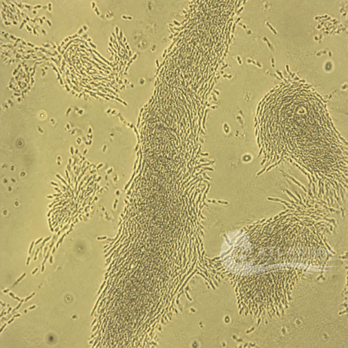
Form, movement and dimensions: Published U. marinum descriptions report a small, rapidly motile ciliate with longitudinal somatic ciliary rows and a caudal cilium. Named studies report different dimensions and do not support a single size range for the entire genus. Fresh relevant material and a measurement scale document an observation, but size, motion or a teardrop outline cannot prove genus, tissue invasion or clinical significance. Microscopy should be interpreted with the relevant sample and diagnostic question.
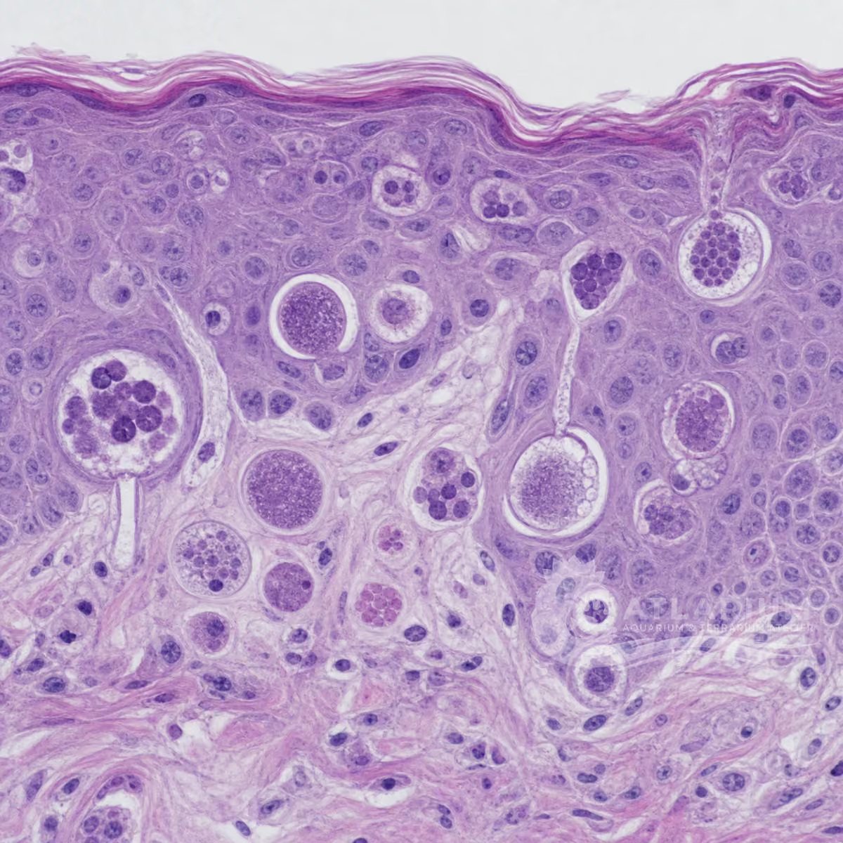
Habitat, host sites and clinical context: Free-living Uronema marinum has been described from organic-matter-associated waters, whereas marine fish-associated reports concern external tissue and sometimes invasive disease. In an imported-ornamental investigation, Uronema sp. was associated with hemorrhagic or ulcerative surface lesions and histologically documented skin, gill, muscle and renal-capsule involvement. Those findings are an outbreak context, not a diagnosis from a lesion, mucus change or free cell.
Ecological role and aquarium relevance: A documented free-living U. marinum can be bacterivorous, while fish-associated Uronema reports show that a ciliate finding can have very different significance in a susceptible host. Its potential to cause disease therefore depends on context. Link a finding to water quality, organic load, affected host, tissue site, burden and diagnostic evidence before inferring risk or cause; do not transfer a free-living observation into a fish-health conclusion.

Observation, culture, and ecological relationships
Where to look and how to sample: In the reported ornamental-fish investigation, diagnostic work used fresh skin and gill scrapings from affected fish, internal examination and histopathology. Lesion, tissue site, fish identity, recent losses and water or husbandry context made the findings interpretable. Nearby water may contain free ciliates without demonstrating invasion, while a negative external sample cannot settle deeper disease. Sampling and handling should meet the diagnostic purpose and professional requirements.
How to observe and identify: Where microscopy is undertaken, examine fresh relevant material at lower power, then use optics to document motile ciliates, movement, burden and tissue change. This is not a diagnostic cutoff. Published U. marinum work combined morphology and molecular identification; outbreak work used histopathology for invasion. Keep conclusions at the supported level when another ciliate, debris, co-infection or noninfectious lesion remains possible; magnification cannot settle agent or tissue depth.
Presence and meaning in an aquarium: A compatible fish case merits assessment of the affected host, tissue site, progression of signs, water quality and organic or husbandry context. In one ornamental-fish study, disease severity was discussed alongside early diagnosis and water-quality management; this does not prove that organic load or any single stressor caused an individual case. A water finding, lesion or response to a change should not be used alone to label Uronema, explain every sign or declare a system safe.
Culture, maintenance or use: No culture, propagation, introduction or biological-control procedure is provided. Published free-living U. marinum studies do not establish an unknown aquarium isolate as suitable starter material, and fish-associated reports do not convert a diagnostic finding into a culture objective. Diagnostic handling needs the relevant sample context and qualified assessment if the result could affect a health or management decision. No general transfer, isolation or equipment protocol is provided.
Documented treatment, control and follow-up: A published investigation of a defined imported ornamental-fish cohort reported formalin baths, a hospital-aquarium phase and follow-up scrapings, with recovery in some fish. It is case-specific evidence, not a general product, dose, duration or treatment protocol. The decision changes with tissue involvement, host tolerance, coexisting disease, authorization and system conditions. Treatment or follow-up therefore requires a confirmed agent or syndrome and qualified veterinary direction.
Risks, biosecurity and limits: The same outbreak literature calls for rapid accurate diagnosis and biosecurity in its ornamental-wholesaler setting, but it supplies no universal quarantine interval, sanitation method, tools rule or prevention protocol. A photo, red lesion or general ciliate label does not establish a contagious outbreak. Progressive ulcers, eye involvement, rapid decline or mortality warrant aquatic-veterinary or laboratory assessment because tissue depth, agent and alternatives affect the response.

Geographic origin and distribution
Origin
Tropical oceans and reefs

Identification and observation data
- Record type
- Genus
- Identification is limited to the genus Uronema. Uronema marinum is a named marine species, but free-living and fish-associated findings do not all establish the same species, host relationship or pathogenic outcome. A moving ciliate, a lesion or a water observation therefore does not identify the genus or diagnose disease. Relevant fresh tissue, morphology, host context and, when the result would affect a health or management decision, specialist or molecular evidence determine the level of identification justified by the evidence.
- Recognition confidence
- Low
- Fresh tissue microscopy can support a ciliate or Uronema-like differential, but small motile forms overlap morphologically and a surface observation does not establish tissue invasion. Published fish investigations combined relevant skin or gill material with histopathology, while a Uronema marinum study used morphology and molecular identification. The conclusion must remain limited to the evidence actually reconciled for the affected host, tissue and system.
- Biological group
- Oligohymenophorea
- Uronema is a ciliate genus in Uronematidae, Philasterida, Scuticociliatia and Oligohymenophorea. Membership in the broad biological grouping of protists and infusoria does not diagnose an organism merely because it is motile. A Uronema marinum reference supports comparison with a named marine taxon, but it cannot turn a freshwater ciliate, an image or a broad scuticociliate finding into that species. Taxonomic confidence depends on the relevant morphological and, when needed, molecular evidence. No order is assigned because the published sources do not establish a well-supported order across the genus Uronema.
- Ecological functions
- Potential pathogen
- The documented ecological role is potential pathogen, not a claim that every Uronema is pathogenic. Free-living Uronema marinum descriptions document bacterivory, whereas fish-associated reports document severe invasive disease in particular hosts and outbreaks. A water observation does not establish disease, and a lesion does not identify the ciliate responsible. Species, tissue site, burden, host condition and diagnostic evidence determine whether clinical relevance is supported.
- Aquarium impact
- Context-dependent
- Impact is context-dependent. A free-living Uronema-like ciliate can be an ecological observation, while a confirmed fish-associated invasive case can be serious. The evidence supports evaluating the relevant sample, host signs, tissue site, water and husbandry context rather than inferring safety or pathology from abundance, a lesion or a general ciliate label. Treatment, control and prevention depend on the confirmed syndrome or agent, system and qualified assessment.
- Culture and management
- Not cultured
- No culture, propagation, introduction or biological-control method is provided. Published free-living Uronema marinum studies and fish-associated disease reports do not make an unknown aquarium isolate appropriate starter material. Diagnostic material and any fish-health response require a purpose, the relevant host or sample context, and qualified veterinary or laboratory assessment where the result is consequential. An observation does not become a culture or management protocol.
- Typical size
- No value supported by reliable evidence
- No genus-wide size value is retained. Published Uronema marinum accounts report different dimensions for named material and preparation can affect what is observed, so a fixed range would falsely imply a genus-level measurement. A measurement scale and fresh, relevant material help document an observed cell, but dimensions alone cannot identify Uronema, prove tissue invasion or establish clinical importance. No genus-wide minimum or maximum size is shown because the published species-level measurements vary by species and method and do not establish a reliable universal size range for Uronema.
- Visible to the naked eye
- No, not as an individual
- Individual Uronema cells are not identifiable with the naked eye. Hemorrhagic or ulcerative surface lesions, altered behavior or other visible fish changes can be compatible with a documented outbreak, but they are not a direct view of a named ciliate. Relevant tissue examination is needed to observe compatible ciliates, and the inference still depends on morphology, tissue site, burden and host context. Visible changes remain open to diagnostic alternatives until those data agree.
- Useful starting magnification
- No universal threshold supported by reliable evidence
- No fixed diagnostic magnification is retained. Published investigations used microscopy with tissue context and, depending on the question, histopathology or molecular identification. The useful setting depends on the sample, objective and optical preparation; magnification alone cannot resolve genus, tissue depth or a treatment decision. When the distinction affects fish health, interpret microscopy together with the affected host, relevant tissue and professional diagnostic evidence.

Links to Aquarium SOS

Microworld ecological network
Links based on documented kinship, food webs, biofilms, and ecological functions.