Columnaris bacterium
Flavobacterium columnare
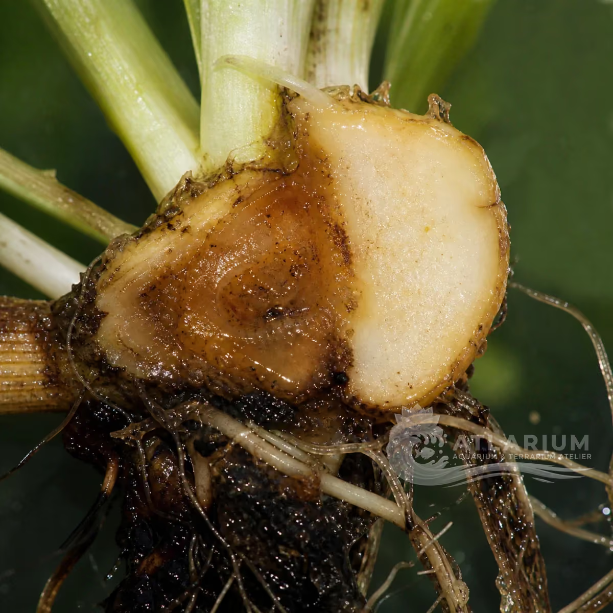
Flavobacterium columnare is a freshwater bacterium associated with columnaris disease in fish. It may produce skin, fin, mouth or gill lesions with yellow-brown, slimy or cotton-like surface material, but appearance alone is not a diagnosis. Use wet mounts only to support a presumptive diagnosis; confirm with suitable culture or molecular testing. Improve water quality, reduce organic loading and avoid traumatic handling while a qualified aquatic veterinarian or laboratory assesses compatible disease.
Freshwater
Flavobacteriia
Species
Medium
Potential pathogen
Potentially harmful
Not cultured
0.3 µm – 10 µm
No, not as an individual
1000× shows cells or aggregates but does not identify the taxon

Identity, habitat, and ecological role

Observation, culture, and ecological relationships

Geographic origin and distribution
Origin

Identification and observation data
- Record type
- Species
- Flavobacterium columnare is the bacterium associated with columnaris disease in freshwater fish. Yellow-brown or cotton-like lesions are not species characters and can have other causes. Use the name only when the appropriate lesion, host, sample and diagnostic evidence agree; a syndrome label, photograph or trade name does not establish the bacterium. Available taxon-specific evidence does not establish a single reliable geographic origin.
- Recognition confidence
- Medium
- A wet mount of affected skin or gill tissue can support a presumptive identification when characteristic bacterial aggregates are present, but it does not establish every look-alike lesion as columnaris. Isolation on appropriate media and molecular assays can confirm the finding. Medium confidence reflects that visual observation alone cannot establish strain identity, causation or treatment without laboratory evidence.
- Biological group
- Flavobacteriia
- F. columnare is a Gram-negative bacterium in the Flavobacteriia, a member of the bacteria. The group does not identify every filament, biofilm or surface growth as this species. Microscopy may show cells or aggregates, while selective culture, staining and molecular tests answer different identity questions. A freshwater finding should not be generalized to marine disease agents.
- Ecological functions
- Potential pathogen
- Its documented role is pathogenic because F. columnare is associated with external columnaris disease in freshwater fish. Disease expression depends on susceptible hosts, skin or gill condition, organic loading, handling stress and other water-quality context. Detecting bacteria in water or a lesion does not alone prove they caused disease; compatible signs and confirmation are needed before a health conclusion is made.
- Aquarium impact
- Potentially harmful
- The potential impact is harmful when compatible fish disease is present, particularly when external skin, fin, mouth or gill lesions progress. It is not a reason to medicate a tank from a photograph. Separate observation, water-quality review, diagnostic sampling and veterinary or laboratory support give safer evidence than reacting to a presumed bacterial name. The same visible lesion can have non-columnaris causes.
- Culture and management
- Not cultured
- This organism is not a home culture. Diagnostic isolation, storage and susceptibility work require qualified laboratory methods, containment and an appropriate clinical purpose. Do not grow, enrich or move unknown lesion material between aquariums. Dedicated tools, careful disposal and separation of affected fish limit spread while the cause is investigated; survival in a sample does not make it suitable for culture or use.
- Typical size
- 0.3 µm – 10 µm
- Cells are commonly described within a 0.3–10 μm working range, but shape, length and grouping vary with strain, sample condition and method. The interval helps set scale for microscopy; it cannot distinguish F. columnare from another bacterium or diagnose columnaris. Note the preparation, microscopic field, host site and scale, then use culture or molecular evidence where species-level identification affects a health decision.
- Visible to the naked eye
- No, not as an individual
- Individual bacterial cells are not reliably visible to the naked eye. A yellow, brown, slimy or cotton-like surface lesion can be conspicuous, but it is host tissue, exudate and mixed material rather than direct evidence of species identity. Visibility of an aggregate cannot identify the bacterium, establish infection severity or justify a treatment; use clinical context and appropriate diagnostic work.
- Useful starting magnification
- 1000× shows cells or aggregates but does not identify the taxon
- At around 1000×, the observer can see cells or characteristic aggregates from affected tissue in a wet mount. These observations may support a presumptive identification but do not provide a definitive species result. Compare several fields and preserve the host site and sample context. Confirmation may require isolation on suitable media, histology or molecular testing. Magnification alone cannot determine antimicrobial susceptibility, systemic spread or the correct intervention.

Links to Aquarium SOS

Microworld ecological network
Links based on documented kinship, food webs, biofilms, and ecological functions.