Aquarium mycobacterium
Mycobacterium marinum
Mycobacterium marinum is an aquatic, acid-fast bacterium associated with granulomatous mycobacteriosis in fish and a possible human skin infection after aquarium exposure. Fish signs are variable and require organism-based confirmation; acid-fast rods are presumptive, not species proof. There is no universal fish treatment to provide here. Use gloves over broken skin, avoid transferring contaminated material, correct or reduce documented husbandry risk factors and seek veterinary, laboratory or medical advice for compatible disease or persistent lesions after exposure.
Freshwater / Brackish / Marine
Actinomycetia
Species
Medium
Potential pathogen
Potentially harmful
Not cultured
0.2 µm – 4 µm
No, not as an individual
1000× shows cells or aggregates but does not identify the taxon

Identity, habitat, and ecological role

Observation, culture, and ecological relationships

Geographic origin and distribution
Origin

Identification and observation data
- Record type
- Species
- Mycobacterium marinum is an aquatic mycobacterium associated with fish mycobacteriosis and a possible human skin infection after aquatic exposure. Granulomas, wasting or a skin lesion are not species characters. Use the name only when host, tissue, exposure history and an organism-based diagnostic result agree; a photograph, a generic disease label or an acid-fast result alone cannot establish this species. Available taxon-specific evidence does not establish a single reliable native geographic origin.
- Recognition confidence
- Medium
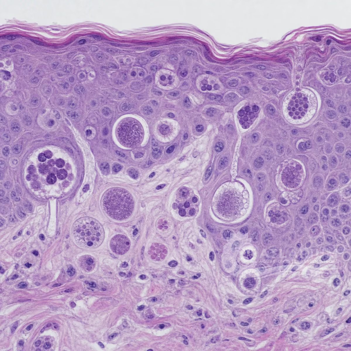
- Acid-fast rods in granulomatous fish tissue can support a presumptive mycobacterial diagnosis, but species confirmation requires isolation and identification or an appropriate molecular method. Disease signs overlap with other disorders and several Mycobacterium species infect fish. Confidence is medium because a visible lesion, wet mount or environmental sample cannot establish species identity, causation, treatment or zoonotic transmission without laboratory evidence.
- Biological group
- Actinomycetia
- M. marinum is an acid-fast bacterium in the Actinomycetia. It is a bacterium, but not every rod, biofilm or granuloma belongs to this species. It has been documented in freshwater, brackish and marine contexts, but a finding in one water type does not identify the organism in another. Microscopy, culture and molecular methods answer different questions and must be interpreted with host and sample context.
- Ecological functions
- Potential pathogen
- Its documented role is pathogenic because M. marinum is associated with granulomatous disease in fish and can be zoonotic after aquatic exposure. Disease expression depends on host, burden, tissue, environmental stress and exposure route. Environmental presence does not prove fish infection, and fish disease does not prove human infection. A health conclusion needs compatible evidence at the relevant host level, with uncertainty retained until organism-based confirmation.
- Aquarium impact
- Potentially harmful
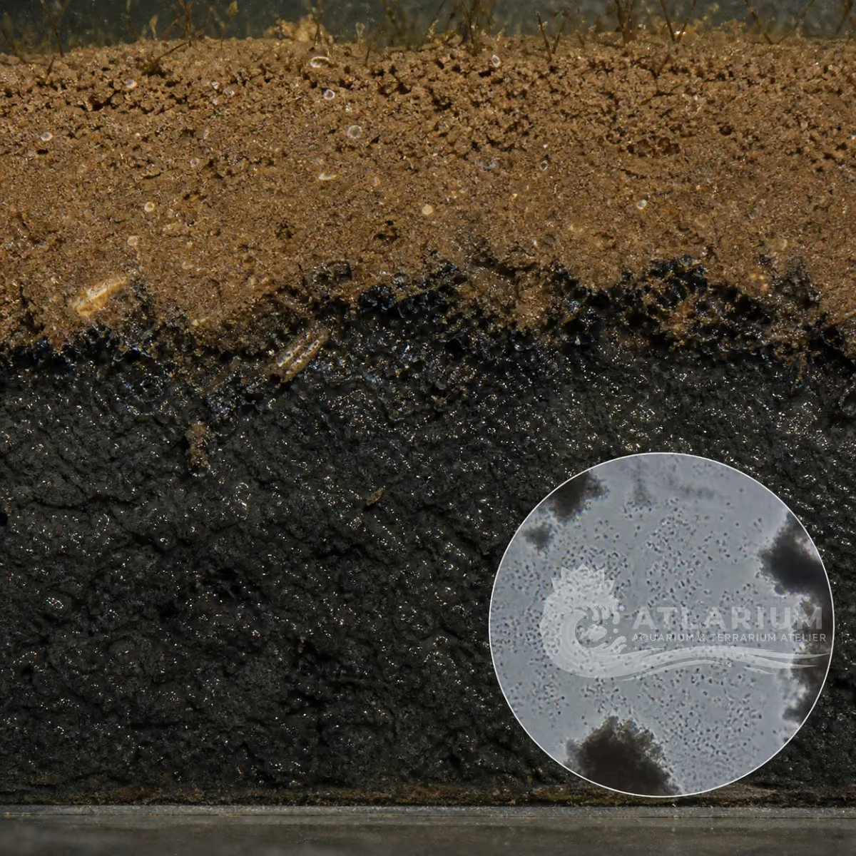
- The potential impact is harmful because compatible fish disease may be chronic and because contact with contaminated aquarium water or material can present a human health risk, particularly through broken skin. It is not a reason to medicate from a photograph or to assume every aquarium is contaminated. Separate fish-health assessment, diagnostic support, careful handling and human exposure precautions are safer than reacting to a presumed bacterial name.
- Culture and management
- Not cultured
- This organism is not a home culture. Diagnostic isolation and identification belong to qualified laboratories with appropriate biosafety procedures. Do not grow, enrich, aerosolize or transfer unknown material from lesions, fish or sediment between aquariums. Dedicated tools, containment and appropriate disposal reduce spread while the cause is investigated. A positive environmental sample or the survival of a culture does not make it suitable for home maintenance or use.
- Typical size
- 0.2 µm – 4 µm
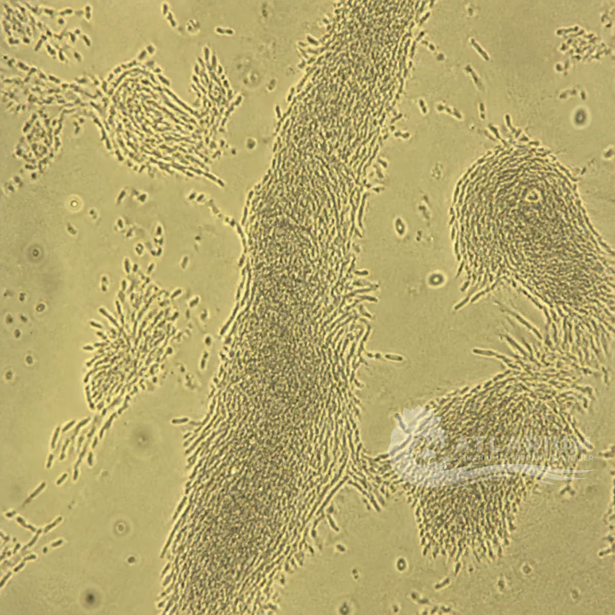
- Cells are commonly described within a 0.2–4 μm working range, but dimensions vary with strain, growth state and preparation. The range sets scale for microscopy; it cannot identify M. marinum, prove infection or distinguish it from another acid-fast mycobacterium. Preserve sample origin, tissue, preparation and scale. Species-level conclusions that affect animal welfare or human exposure require culture identification or another validated organism-based method.
- Visible to the naked eye
- No, not as an individual
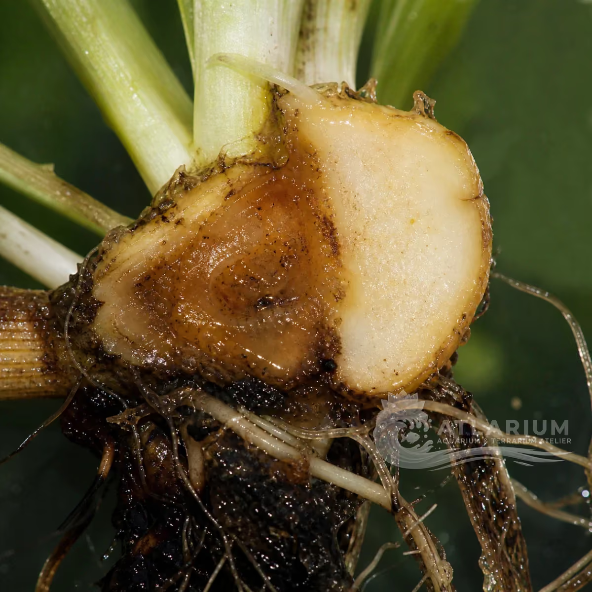
- Individual cells are not reliably visible to the naked eye. Fish nodules, ulcers, wasting or tissue changes may be visible but are host responses or mixed material, not direct evidence of species identity. A visible skin lesion in a person likewise needs medical assessment rather than aquarium self-diagnosis. Appearance cannot establish source, severity, transmissibility or a treatment. Keep observation, diagnosis and exposure response separate.
- Useful starting magnification
- 1000× shows cells or aggregates but does not identify the taxon
- At around 1000× with appropriate staining, the observer may see acid-fast rods in granulomatous material. This finding can support a presumptive mycobacterial diagnosis but cannot confirm M. marinum, determine susceptibility, prove systemic disease or diagnose a human skin lesion. Compare several fields and retain tissue and exposure context. Confirmation may require culture under suitable conditions, identification and molecular work by qualified veterinary, medical or diagnostic laboratories.

Links to Aquarium SOS

Microworld ecological network
Links based on documented kinship, food webs, biofilms, and ecological functions.