Trichodina disc ciliates
Trichodina
Trichodina is a genus of disc-shaped ciliates found on the skin, gills and fins of aquatic hosts. Low burdens can be contextual, whereas heavy infestation may cause excess mucus, flashing, respiratory effort and loss of condition. A circular ciliate on one slide is not a species diagnosis or proof of disease. Confirm relevant wet mounts, assess burden and host signs, correct crowding and sanitation, and use chemical treatment only under appropriate fish-health guidance.
Water type
Freshwater / Brackish / Marine
Biological group
Oligohymenophorea
Record type
Genus
Recognition confidence
Low
Ecological functions
Parasite, Potential pathogen
Aquarium impact
Context-dependent
Culture and management
Not cultured
Typical size
20 µm – 100 µm
Visible to the naked eye
No, not as an individual
Useful starting magnification
400× to begin observation

Identity, habitat, and ecological role
Identity and taxonomic resolution: Trichodina is retained at genus level because species differ in host range, salinity and clinical effect. These discoid ciliates can be ectocommensal or parasitic on fish and other aquatic hosts. Outline or spinning movement can inform a differential, but mucus, debris and related trichodinids can resemble the organism. A defensible finding combines fresh material, attachment site, diagnostic structures and host context. Do not turn a genus finding into species certainty or cause.
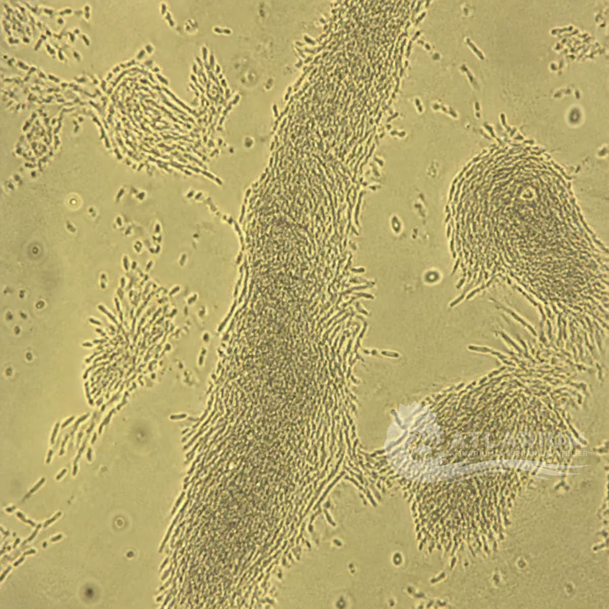
Form, attachment and dimensions: Trichodina forms a flattened disc with a characteristic adhesive apparatus and ciliary movement; it is often seen rotating on fresh skin, fin or gill material. The 20–100 μm working range varies by species and preparation. Observe several intact individuals with a scale and note their site. Size, rotation and disc shape provide supporting evidence but cannot independently establish species, burden or pathogenicity.
Host sites and clinical context: Relevant sites include skin, gills and fins in freshwater, brackish or marine fish systems. Heavy burdens can be associated with excess mucus, flashing, rapid respiration, piping, gill damage and loss of condition, while an external finding without compatible signs may have different meaning. Species, salinity, tissue site, parasite number, co-infections and water quality influence outcome. Affected-tissue wet mounts are necessary; visible mucus cannot identify Trichodina.
Burden, husbandry and relevance: Trichodinids are often associated with overcrowding, poor sanitation and high organic material. Note stocking, feeding, water, detritus, filters, transfers and fish behavior before linking a ciliate to disease. Cleaning the system and reducing conditions that support organic build-up are part of control, not an optional add-on to medication. One low count does not prove a tank outbreak; treatment response does not settle every ciliate identity.

Observation, culture, and ecological relationships
Where to look and how to sample: Sample fresh mucus, skin, fin and gill material from fish with compatible signs, and note the species, tank, date, salinity, water quality, stocking, feeding and recent changes. Prepare wet mounts promptly and retain the exact tissue-site information. Water samples may show free material but do not replace examination of the host surface. One negative site cannot exclude a focal burden, while a positive sample must not be generalized to every fish or tank.
How to observe and identify: Scan fresh material at low power, then use about 400× to compare several small rotating disc ciliates, their attachment structures and associated tissue changes. Note counts consistently and distinguish likely Trichodina from debris, mucus and related trichodinids. Wet mounts support identification at genus level when morphology, tissue and signs agree. Species identity, unusual severity or uncertain lesions may require specialist microscopy, staining, molecular work or veterinary assessment.
Immediate management and containment: When compatible signs and wet-mount evidence support a case, assess host context before selecting response. Review oxygenation, ammonia, nitrite, temperature, salinity, stocking, feeding, filtration and organic debris. Associations with crowding or poor sanitation guide that context, but do not supply a universal isolation, sharing, handling or sanitation procedure. These considerations do not replace diagnosis, and a water or husbandry finding alone does not establish Trichodina as the cause.
Treatment and professional limits: Veterinary references list formalin or copper treatment for confirmed external trichodinid disease; choice depends on freshwater or marine context. Formalin is toxic and can affect biological filtration; copper suitability and hazards need evaluation for the diagnosed host, water and formulation. No dose, duration or universal protocol is provided. Use only an authorised label or aquatic-veterinary direction after confirmation, and monitor the system and susceptible non-target organisms.
Control, follow-up and prevention: Reassess fish signs, tissue wet mounts, organic material and water quality after management or treatment as directed by a clinician. Persistent problems warrant review of burden, host, co-infections, salinity, filtration and crowding. For a diagnosed case, those factors inform context-bound management; no universal quarantine interval, dedicated-tools rule, transfer ban or preventive-medication procedure follows. Treatment response does not determine identity or explain every cause.
Risks, biosecurity and limits: Do not diagnose Trichodina, contagion or a treatment need from a photo alone. Follow product safety information only if a professional directs medication after confirmation. Seek aquatic-veterinary or laboratory help for rapid respiratory decline, losses, gill disease or uncertain identifications. The genus, burden and clinical role remain conditional until organism, tissue, host signs and alternatives have been reconciled; no universal home containment or transfer procedure is established.

Geographic origin and distribution
Origin
North America

Identification and observation data
- Record type
- Genus
- Identification is limited to the genus Trichodina. The genus includes multiple species that differ in host range, salinity and clinical significance. A spinning disc in a wet mount or a circular image does not establish a species, a disease outbreak or a specific host relationship. Stronger identification requires fresh material, the relevant tissue site and diagnostic structures; consequential cases may need specialist morphology or molecular work.
- Recognition confidence
- Low
- Fresh wet mounts can support identification as Trichodina when several intact rotating discs and their attachment structures are seen on relevant host tissue. Mucus, debris and related trichodinids can look similar, so a single image is not enough. Note scale, tissue site, salinity and burden. Species certainty and causal attribution require more evidence, particularly where gill disease, mortality or mixed infection is present; alternatives must be reconciled before a consequential treatment decision.
- Biological group
- Oligohymenophorea
- Trichodina is a ciliate within Oligohymenophorea. It belongs to the broad biological grouping of protists and infusoria, but that grouping does not make all small aquatic cells equivalent to Trichodina. Its cilia and adhesive disc explain its wet-mount appearance and external association with hosts. Taxonomic context must not merge Trichodina with other trichodinids or turn a disc-shaped particle into a parasite diagnosis without relevant morphology and host context. Published taxon-specific evidence supports the genus-level diagnostic context, but establishes neither a reliable order nor a single natural geographic origin for Trichodina.
- Ecological functions
- Parasite, Potential pathogen
- Trichodina can act as a parasite and potential pathogen. Some trichodinids may be ectocommensal at low burden, while heavy infestation on skin or gills can cause irritation and tissue damage. Clinical effect depends on species, number, host condition, salinity, organic load and co-infections. Burden-aware sampling and husbandry correction are therefore important, but detection alone does not show that a ciliate is harmful or sufficient to explain every sign.
- Aquarium impact
- Context-dependent
- The aquarium impact is context-dependent. Heavy compatible burdens can compromise fish condition and breathing, while a low-level or environmental finding may have a different meaning. Relevant wet mounts, water-quality review and host context come before any consequential treatment decision. Do not use visual resemblance as a treatment trigger, and do not assume that a chemical response identifies the organism or corrects overcrowding and organic accumulation.
- Culture and management
- Not cultured
- No culture or propagation method is provided because the cited evidence concerns external fish disease, not maintenance of a Trichodina isolate. An incidental finding may prompt a genus-level diagnostic investigation, but does not establish a home culture or any rule for exchange, transfer or containment. In a diagnostically supported case, handling and any husbandry decision depend on the host, system, water context and aquatic-veterinary plan.
- Typical size
- 20 µm – 100 µm
- The 20–100 μm range is a broad guide for disc-shaped ciliates within this genus. Size varies by species, viewing angle, tissue site and preparation. Measure several intact individuals with a calibrated scale and note the host and salinity context. Dimensions alone cannot distinguish Trichodina from related trichodinids, prove burden or determine whether the observed organism is clinically relevant.
- Visible to the naked eye
- No, not as an individual
- Individual Trichodina cells are not identifiable with the naked eye. Excess mucus, flashing, poor condition or rapid breathing can be visible host clues, but none is a direct view of the ciliate. Relevant fresh wet mounts are required to observe rotating discs and their structures. Diagnostic confidence still depends on tissue site, burden, host signs and exclusion of alternatives, not on the appearance of one fish or photograph.
- Useful starting magnification
- 400× to begin observation
- Start at low power to find representative skin, fin or gill material, then use around 400× to inspect rotating disc ciliates, attachment structures and associated tissue changes across several fields. Capture scale and site information. This supports identification at genus level but does not identify every species or establish a clinical role. Specialist microscopy, staining or molecular work may be needed before consequential treatment decisions.

Links to Aquarium SOS

Microworld ecological network
Links based on documented kinship, food webs, biofilms, and ecological functions.