Freshwater ich ciliate
Ichthyophthirius multifiliis
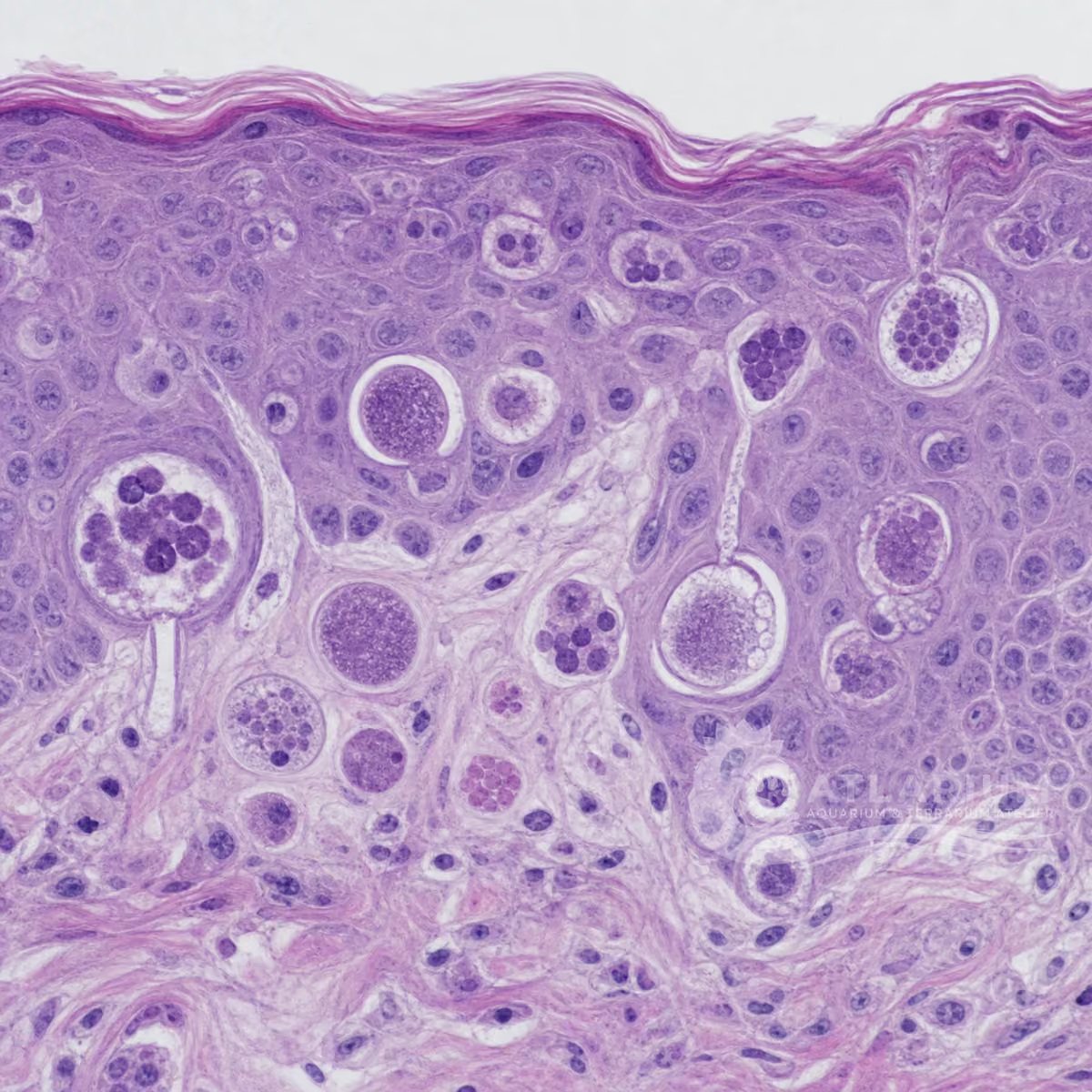
Ichthyophthirius multifiliis is a freshwater ciliate that causes ich only when confirmed in compatible fish tissue. Its direct cycle has on-host trophonts, environmental tomonts and infective theronts, so visible white spots are neither the whole lifecycle nor a diagnosis. Examine fresh preparations from skin, fins and gills microscopically alongside the fish’s signs and system context; treatment, control and prevention must follow the confirmed stage and case.
Water type
Freshwater
Biological group
Oligohymenophorea
Record type
Species
Recognition confidence
Medium
Ecological functions
Parasite, Potential pathogen
Aquarium impact
Potentially harmful
Culture and management
Not cultured
Typical size
50 µm – 1,000 µm
Visible to the naked eye
Yes, as an individual
Useful starting magnification
100× to begin observation
Identity, habitat, and ecological role
Identity and taxonomic resolution: Ichthyophthirius multifiliis is a species-level freshwater fish ciliate, not a label for every white mark on a fish. White spots can be compatible with ich but overlap with other lesions, and gill-dominant cases may have no visible spots. A defensible result joins host, tissue, wet mount, compatible morphology and life stage. When these observations are incomplete, keep ich among the differential diagnoses or seek qualified laboratory confirmation; suspicion alone is not confirmation.
Morphology and dimensions: The 50–1,000 μm guide spans substantially different lifecycle stages and is an observation range, not a stand-alone diagnosis. On-host trophonts are larger than free-swimming theronts; tissue thickness, compression and sample condition can alter what is seen. A wet mount should preserve sample site, scale, magnification and movement. A rounded ciliate and compatible macronucleus can support identification, but size, a photograph or one field alone cannot distinguish every look-alike.
Habitat and microhabitat: This freshwater parasite occupies different microhabitats during one direct lifecycle. The trophont feeds in fish skin or gill epithelium; the tomont leaves the host and reproduces in the environment; theronts then seek a compatible fish. Host tissue, water, substrate, filter material, plants and equipment therefore have different relevance to observation and transfer. A finding at one site does not demonstrate infection throughout a tank.
Food-web role and aquarium meaning: I. multifiliis is an obligate fish parasite and potential pathogen, not a cleanup organism or live food. The lifecycle can amplify under compatible host conditions, while visible burden and outcome depend on tissue site, fish condition and stage. A white mark, respiratory sign or environmental cell is not proof of ich. Keep compatible signs, confirmed parasite, husbandry factors and alternative diagnoses distinct when assessing urgency or system risk.

Observation, culture, and ecological relationships
Where and how to sample: Collect fresh skin mucus, fin and gill material from fish with compatible signs, keeping these sites separate. Note the host, date, system, recent transfers, temperature and measured water conditions; the method and site are part of the result. Use clean dedicated tools and avoid moving water or mucus between aquariums. A negative preparation does not prove absence when the wrong tissue, stage or timing was sampled, so preserve the limitation rather than declaring a system free of infection.
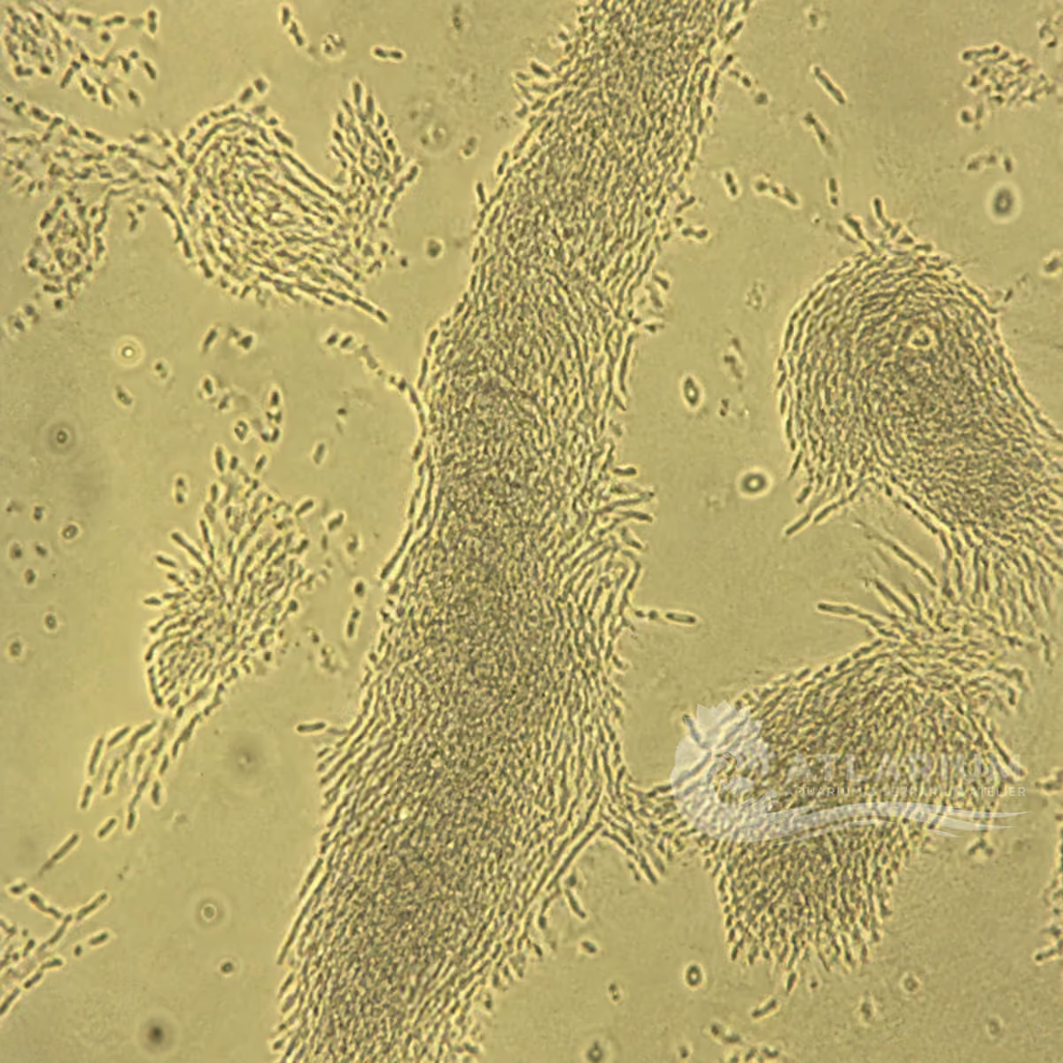
How to observe and identify: Examine a wet mount first at lower power, then increase magnification to assess ciliation, movement and any visible horseshoe- or C-shaped macronucleus. Compare multiple fields and, when warranted, skin, fin and gill samples. Mature trophonts can be easier to recognize than immature theronts, which overlap with other small organisms. Do not name a parasite solely from a photograph or a white spot.
Reading a finding in the aquarium: Interpret microscopy with fish behaviour, respiratory effort, abundance, tissue location and water quality. White spots may be absent when gills are important, and the same visible sign can have non-ich causes. Stabilise basic husbandry and document the trend while the differential is assessed; do not treat an image as proof of contagion. Severe respiratory signs, spread or mortality justify prompt fish-health support.
Culture, containment and transfer: This parasite is not a home culture organism and no propagation protocol is provided. Do not keep, transfer, aerosolise or intentionally multiply suspect material. Isolate or restrict movement of affected fish where appropriate, use dedicated equipment, prevent water and mucus transfer, and dispose of material responsibly. Diagnostic isolation and experimental maintenance belong to qualified fish-health facilities.
Treatment, control and prevention: Treatment is not a universal product, dose or duration: on-host trophonts and protected cyst stages are not equivalent to susceptible free-swimming theronts. Confirm the diagnosis, identify lifecycle timing and obtain fish-health advice before selecting an intervention. Control and prevention include limiting transfers, quarantine, dedicated equipment, observation and water-quality management. These measures do not authorize medication without host, system, legal and product context.
Limits, documentation and specialist referral: Keep host identity, tissue site, signs, dates, water values, images, magnification and actions together in the case notes. Do not infer treatment success merely because spots change, since stage transitions can alter visibility. Escalate progressive disease, high losses, uncertain identification or decisions affecting animal movement to an aquatic veterinarian or qualified laboratory. These notes do not replace case-specific examination, medicine labels or local requirements.
Identification and observation data
- Record type
- Species
- Ichthyophthirius multifiliis is a freshwater ciliate associated with ich when confirmed in compatible fish tissue. The name and visible white spots are not a diagnosis: other lesions can look similar and gill-dominant disease may lack external spots. A tissue wet mount, compatible morphology and case context support the species-level interpretation more strongly than a photograph or an aquarium-wide assumption. Published evidence documents this species in freshwater-fish disease contexts worldwide, but does not establish one well-supported natural geographic origin.
- Recognition confidence
- Medium
- Confidence is high only when skin, fin or gill wet mounts show a compatible ciliate in the right host and clinical context. Large on-host trophonts may show slow rolling movement, ciliation and a horseshoe- or C-shaped macronucleus; immature theronts are smaller and overlap with look-alikes. Sample site and lifecycle stage matter. Do not diagnose ich from white spots alone or exclude it from a gill-only presentation without microscopy.
- Biological group
- Oligohymenophorea
- Ichthyophthirius multifiliis is a freshwater ciliate in Oligohymenophorea and Ichthyophthiriidae. Its ciliate placement among protists and infusoria does not replace the exact species-level fish-parasite identity. It must not be confused with Cryptocaryon irritans, a marine parasite associated with a superficially similar syndrome but a different system and taxonomic context. Keep freshwater and marine disease claims separate.
- Ecological functions
- Parasite, Potential pathogen
- It is an obligate fish parasite and potential pathogen. The trophont feeds in host epithelium, the tomont reproduces after leaving the host and theronts seek fish, so this direct lifecycle can amplify and affect skin or gills. It does not mean every white mark is ich or that visible burden alone predicts outcome. Tissue confirmation, stage, host condition and compatible signs determine how a finding should be interpreted.
- Aquarium impact
- Potentially harmful
- A confirmed infection can be serious, particularly when gills are affected, but white spots, clamped fins, lethargy and respiratory signs are compatible rather than exclusive findings. Treat a suspect case as urgent for documentation, diagnosis, isolation where appropriate and support of safe water conditions. Keep observed signs distinct from confirmation. The relevant risk follows the host-associated lifecycle and potential transfer, not the mere presence of free-water cells.
- Culture and management
- Not cultured
- I. multifiliis is not a home culture organism. It can move with infected fish and contaminated water, substrate, plants, structures or equipment, so suspect material must not be maintained or transferred. Use dedicated tools, quarantine and containment around a diagnostic case. Qualified fish-health facilities, not an aquarium hobby protocol, are the appropriate setting for any diagnostic isolation or experimental maintenance of this fish parasite.
- Typical size
- 50 µm – 1,000 µm
- The reported 50–1,000 μm range covers different lifecycle stages; mature trophonts are commonly described around 0.5–1.0 mm, while free-swimming theronts are much smaller. Size must be tied to stage, tissue site and a scale and cannot identify the organism by itself. Thick, compressed or deteriorated tissue can hide the macronucleus and distort apparent dimensions, so preserve preparation and measurement limits with the observation.
- Visible to the naked eye
- Yes, as an individual
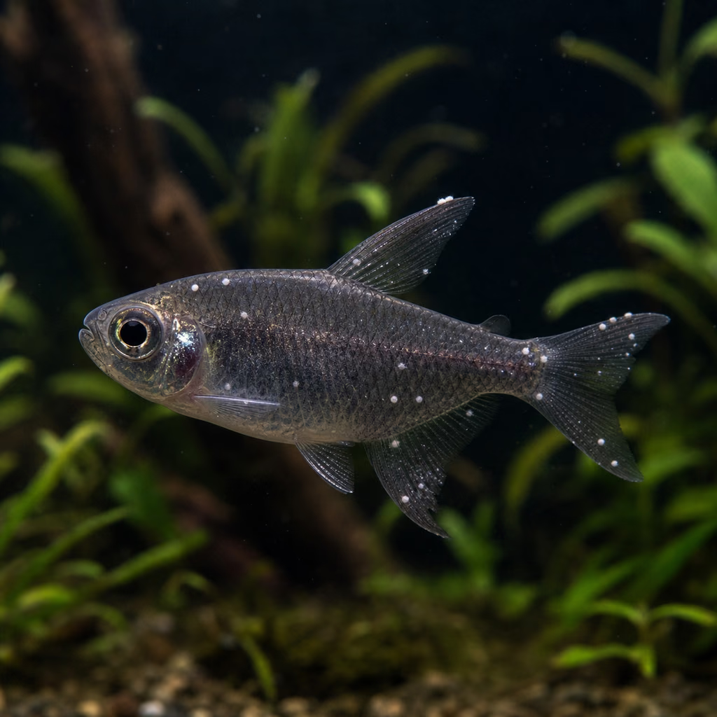
- Mature trophonts in skin or fins can appear as white nodules visible to the naked eye, but that sign is neither always present nor unique to ich. Gill-dominant infection may show no external spots. Visibility therefore supports suspicion rather than identification. Microscopic examination of fresh skin, fin and gill preparations is needed to see the parasite in context, distinguish a compatible finding from look-alikes and avoid treating a visible mark as a diagnosis.
- Useful starting magnification
- 100× to begin observation
- Start around 40–100× for mature trophonts in wet mounts, then increase power to inspect ciliation and the macronucleus. Examine skin, fin and gill material separately because distribution differs among sites and stages. Immature theronts can be difficult to distinguish, so one microscopic field or stage is insufficient. Microscopy supports diagnosis; treatment decisions also require the fish, water, lifecycle and system context rather than magnification alone.
Links to Aquarium SOS
Microworld ecological network
Links based on documented kinship, food webs, biofilms, and ecological functions.