Aquatic-plant soft-rot bacteria
Più famiglie batteriche
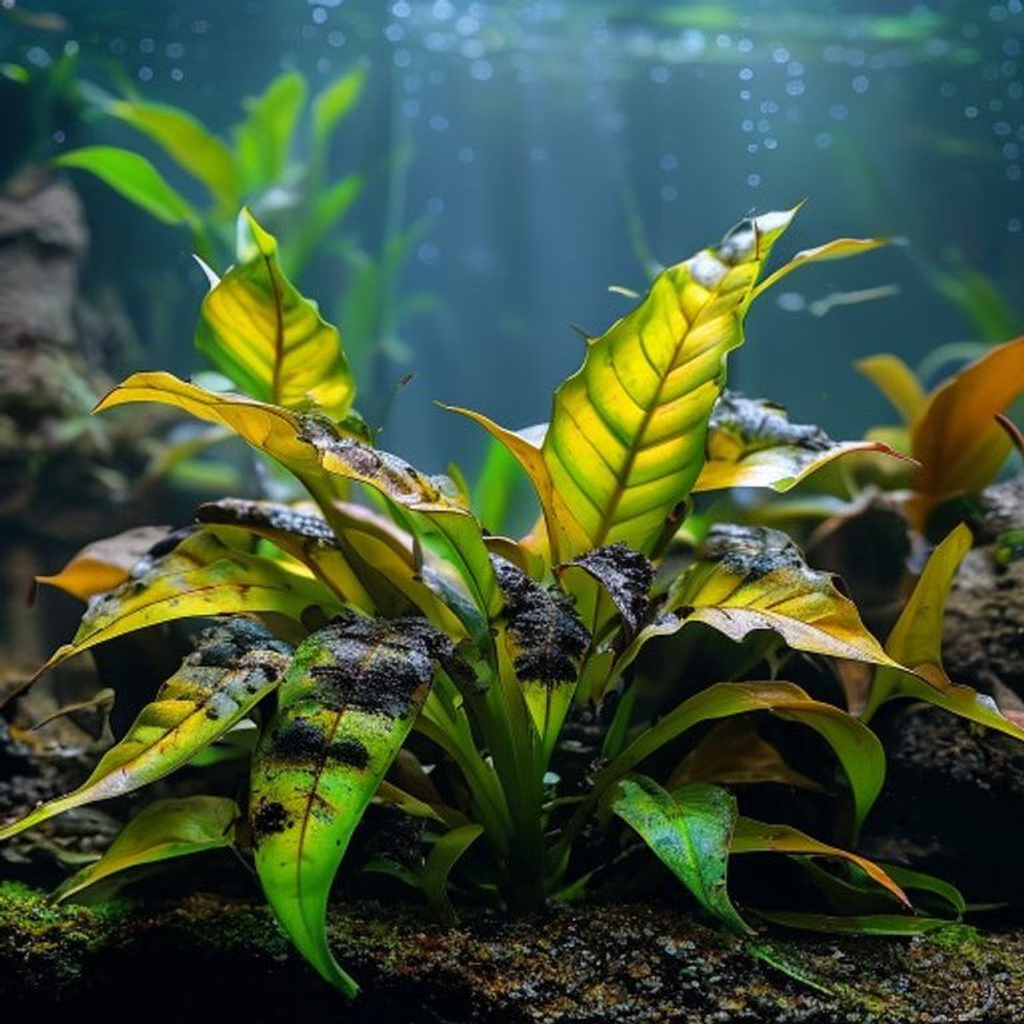
Soft rot is a plant-tissue syndrome, not a species diagnosis. Pectobacterium and Dickeya include documented plant pathogens, but a melting aquatic plant can also reflect transport injury, unsuitable emersion-to-submersion transition, nutrient stress or another disease. Water-soaked, soft and collapsing tissue should prompt isolation, documentation and plant-specific diagnosis; it does not justify antibiotics or a universal aquarium chemical treatment.
Freshwater / Brackish
Più famiglie batteriche
Functional group
Low
Decomposer, Potential pathogen
Context-dependent
Not cultured
0.5 µm – 5 µm
Varies by taxon or life stage
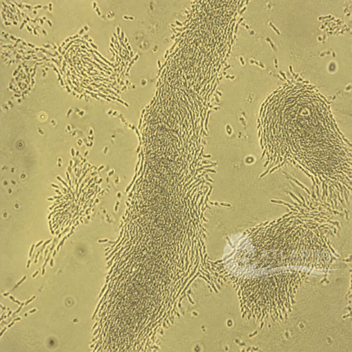
1000× shows cells or aggregates but does not identify the taxon

Identity, habitat, and ecological role

Observation, culture, and ecological relationships

Geographic origin and distribution
Origin

Identification and observation data
- Record type
- Functional group
- This is a functional syndrome entry for bacteria associated with aquatic-plant soft rot, not a species or genus. Pectobacterium and Dickeya are important documented examples, but the same visible collapse can result from another microbe or a noninfectious cultivation problem. The label organizes a cautious investigation of macerated tissue; it cannot name a bacterium, host range, transmission route or treatment without plant-specific diagnostic evidence.
- Recognition confidence
- Low
- Confidence is low because water-soaked, soft or collapsing plant tissue is a syndrome rather than a bacterial character. Ordinary microscopy may show cells but cannot establish a genus, virulence or causation, and bacteria frequently colonize tissue after another injury. Preserve a fresh lesion margin and plant context, compare noninfectious explanations and use culture or molecular plant-diagnostic methods when a precise cause would change management or plant movement.
- Biological group
- Più famiglie batteriche
- The aquarium-facing group is bacteria and archaea, while this entry deliberately spans multiple possible bacterial families instead of declaring a taxon. Pectobacterium and Dickeya are examples of soft-rot plant pathogens, not automatic identities for every aquatic-plant lesion. The group label guides careful sampling and differential diagnosis; it does not turn cell shape, biofilm or a rotting leaf into proof of pathogenic bacteria.
- Ecological functions
- Decomposer, Potential pathogen
- Decomposer and pathogen are distinct contextual roles. Bacteria can recycle already dead plant material, whereas a confirmed soft-rot pathogen can initiate or intensify tissue maceration under suitable host and environmental conditions. Finding bacteria on decay does not show which role came first. Record symptom progression, plant history, lesion margin and environmental changes before assigning responsibility, and do not generalize a plant syndrome into a risk for aquarium animals.
- Aquarium impact
- Context-dependent
- Impact is context dependent. Rapid plant collapse can increase organic load and destabilize a small system, while a localized lesion may be confined to one damaged plant. The syndrome itself does not establish contagion, a fish disease or the need to medicate water. Isolate affected material, assess husbandry and identify the plant and causal process before choosing a response. Chemical intervention without confirmation can harm plants, invertebrates, fish and useful microbes.
- Culture and management
- Not cultured
- This group is not cultured at home because it represents possible plant pathogens and opportunistic colonizers rather than an identified safe strain. Diagnostic isolation belongs to qualified laboratories using appropriate containment and controls. Aquarium management should focus on healthy plant conditions, removal of macerated tissue and prevention of uncontrolled transfer. Do not grow, share or deliberately spread unknown bacteria from a rotting plant in order to test a diagnosis.
- Typical size
- 0.5 µm – 5 µm
- The displayed 0.5–5 µm range is a broad guide to bacterial cell dimensions, not the size of a lesion, biofilm or taxonomic boundary. Cell size overlaps widely across harmless and pathogenic bacteria and varies with strain, growth state and preparation. Measure only with appropriate microscopy and a scale. A cell within this range cannot be assigned to Pectobacterium, Dickeya or any causal role without validated diagnostic evidence.
- Visible to the naked eye
- Varies by taxon or life stage
- Individual bacterial cells in this group are not visible to the naked eye. What an aquarist can see is the plant response: water-soaked areas, softening, discoloration, collapse, debris or possibly odour. Those signs are useful for documenting a syndrome but not for identifying a bacterium. Photograph the intact plant and lesion margin, record the time course and retain a fresh sample if qualified diagnostic assessment is needed.
- Useful starting magnification
- 1000× shows cells or aggregates but does not identify the taxon
- About 1000× total magnification with appropriate preparation may reveal bacterial cells or aggregates, but it cannot confirm a genus, pathogenicity or disease cause. Staining, isolation, biochemical methods and molecular assays are required for a defensible plant-pathogen identification. Record the preparation, scale, lesion margin and plant history. The useful starting magnification is therefore for observation, not a threshold that turns a microscopic image into a diagnosis.

Links to Aquarium SOS
Microworld ecological network
Links based on documented kinship, food webs, biofilms, and ecological functions.