Diseases
Database of fish pathologies, symptoms, and treatments.
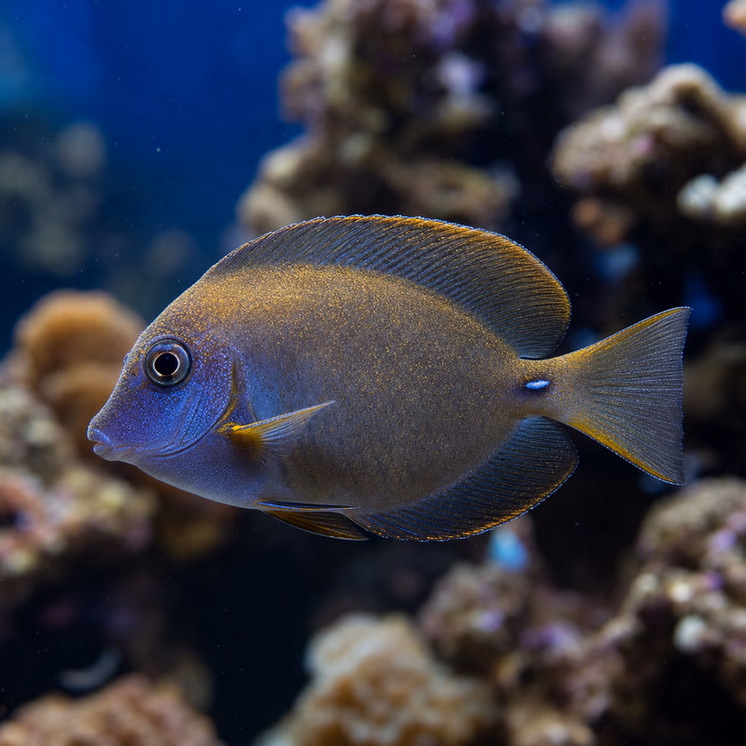
Black Ich / Tang Disease
Paravortex spp.
Pathophysiological Overview This condition represents a highly virulent, systemic pathology deeply rooted in acute immunological distress. Microscopic analysis typically reveals profound tissue necrosis and cellular degradation at the epithelial level. Epidemiological Factors Transmission is significantly enhanced in closed-system aquatic or terrarium environments with elevated bioloads. Pathogenic proliferation is logarithmic once the primary host defenses are breached. Lifecycle & Virulence The disease progression is characterized by a rapid onset, leveraging host stress pathways to bypass primary mucus or epidermal barriers.
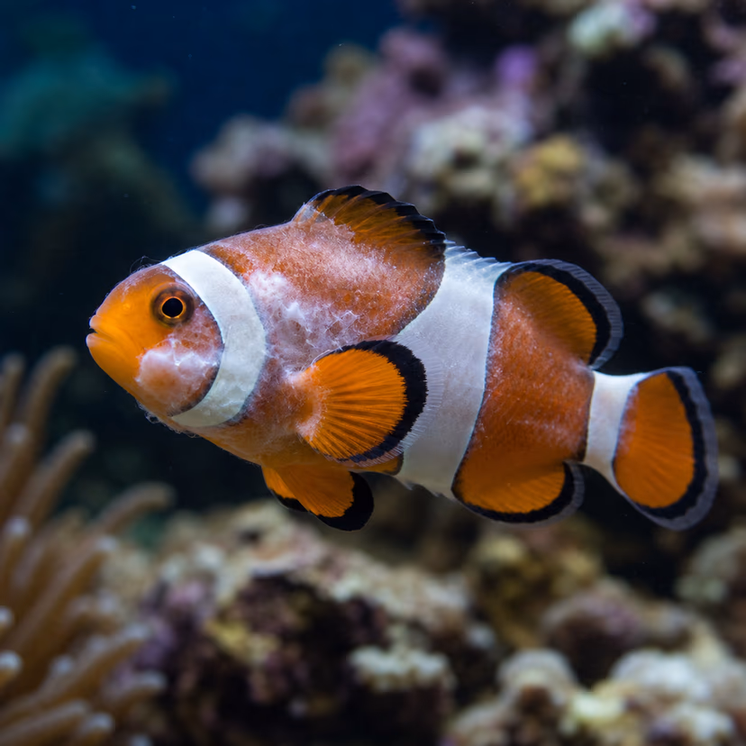
Brooklynella (Clownfish Disease)
Brooklynella hostilis
Ciliate Pathology and Cytotoxicity Brooklynella hostilis is an aggressive, bean-shaped, ciliated protozoan notorious for decimating captive marine populations, earning the colloquial moniker 'Clownfish Disease' due to its disproportionate lethality in Amphiprioninae. Unlike Cryptocaryon or Amyloodinium, Brooklynella reproduces via simple binary fission directly on the host, bypassing an environmental encysted stage. This direct reproductive cycle allows the parasite population to multiply exponentially within hours on a single host. The ciliate feeds ruthlessly on the epidermal cells and blood cells, causing intense irritation that triggers the host’s goblet cells to secrete massive, lethal quantities of opaque mucus.
Camallanus Worms
Camallanus spp.
Nematode Biology and Parasitism Camallanus cotti and related species are formidable, blood-feeding nematode parasites that aggressively infect the lower intestines and rectum of freshwater teleosts, particularly livebearers (Poeciliidae), cichlids, and anabantoids. Unlike many parasitic nematodes, Camallanus features a direct or facultative indirect life cycle. Viviparous females expel fully formed, free-living, first-stage larvae ($L_1$) directly into the water column. These larvae can be ingested by intermediate hosts (copepods or crustacean larvae) or ingested directly by a susceptible fish. Upon ingestion, the larvae molt and migrate to the intestine, where they utilize a heavily sclerotized, jaw-like buccal capsule to anchor deep into the mucosal lining, feeding continuously on the host's blood.

Chytrid Fungus
Batrachochytrium dendrobatidis
Pathophysiological Overview This condition represents a highly virulent, systemic pathology deeply rooted in acute immunological distress. Microscopic analysis typically reveals profound tissue necrosis and cellular degradation at the epithelial level. Epidemiological Factors Transmission is significantly enhanced in closed-system aquatic or terrarium environments with elevated bioloads. Pathogenic proliferation is logarithmic once the primary host defenses are breached. Lifecycle & Virulence The disease progression is characterized by a rapid onset, leveraging host stress pathways to bypass primary mucus or epidermal barriers.
Columnaris (Cotton Wool Disease)
Flavobacterium columnare
Microbiology of Flavobacterium columnare Columnaris is a highly contagious and acutely fatal disease caused by the Gram-negative, aerobic, rod-shaped bacterium Flavobacterium columnare. The disease is often misdiagnosed as a fungal infection due to its filamentous, cotton-like appearance on the epidermis. F. columnare possesses a robust affinity for gill and skin tissue, where it utilizes specialized gliding motility to colonize and form dense biofilms. The bacterium releases tissue-degrading enzymes, notably chondroitinases and proteases, which digest the epithelial and dermal layers, leading to rapid and widespread necrosis. Epidemiological Dynamics The virulence of F. columnare is highly temperature-dependent, with explosive outbreaks typically occurring when water temperatures exceed $25^{\circ}C$. High organic loads and elevated pH further exacerbate the pathogenesis, accelerating the bacterial division rate and biofilm maturation.
Dropsy
Aeromonas spp.
Pathophysiology of Dropsy Dropsy is not a singular pathogen but rather a clinical manifestation of severe systemic disease, primarily characterized by pronounced edema and ascites. The condition arises from a catastrophic failure of the teleost osmoregulatory system, specifically involving renal or hepatic dysfunction. In freshwater environments, fish are hyperosmotic relative to their surroundings, necessitating constant water excretion via the kidneys. When internal organs are compromised—frequently by Gram-negative bacteria such as Aeromonas hydrophila or Pseudomonas fluorescens—fluid accumulation rapidly outpaces excretion. This internal hydrostatic pressure forces the scales outward, resulting in the pathognomonic 'pinecone' appearance. Etiological Factors The etiology is multifactorial but overwhelmingly bacterial in origin. Opportunistic pathogens colonize the host following an immunosuppressive event, often mediated by environmental stressors such as chronic ammonia toxicity, sudden thermal shifts, or prolonged poor water quality. Virulence factors of Aeromonas species include aerolysins, hemolysins, and proteases that cause severe tissue necrosis, particularly in the nephronic tubules and hepatic parenchyma, culminating in multi-organ failure.
Epistylis
Epistylis spp.
Protozoology and Symbiotic Mechanics Epistylis is a genus of peritrichous, ciliated protozoans that are fundamentally distinct from the parasitic ciliate Ichthyophthirius multifiliis (Ich). While often visually confused with Ich, Epistylis organisms are structurally unique: they are colonial and anchor themselves to the host’s epithelium via a rigid, non-contractile, calcified stalk. From the apex of this stalk, a bell-shaped zooid utilizes its cilia to filter-feed on waterborne bacteria and organic detritus. Crucially, Epistylis is primarily an epibiont (a commensal organism) rather than an obligate tissue parasite. However, the calcified stalk pierces the host's epidermal layer. In water heavily laden with organic pollutants and high bacterial counts (specifically Aeromonas spp.), these puncture wounds act as direct vectors for lethal systemic bacterial septicaemia. The morbidity associated with Epistylis is almost exclusively due to these secondary bacterial infections.

Fin Rot
Pseudomonas spp., Aeromonas spp.
Bacterial Pathophysiology Fin Rot is not a singular pathogen, but rather an opportunistic polymicrobial infection primarily caused by ubiquitous, gram-negative, motile rod-shaped bacteria—most notably Aeromonas hydrophila, Pseudomonas fluorescens, and occasionally Flexibacter species. Mechanisms of Virulence: These pathogens are universally present in all aquatic environments. They rely entirely on pre-existing physical trauma or severe immunosuppression in the host to breach the integumentary barrier. Once attached, they secrete powerful extracellular proteases and hemolysins that rapidly digest the structural collagen and soft connective tissue between the fin rays (lepidotrichia).
Fish Tuberculosis (Fish TB)
Mycobacterium marinum
Pathobiology of Piscine Mycobacteriosis Piscine Mycobacteriosis, colloquially termed 'Fish TB', is an insidious, chronic, and largely untreatable bacterial infection predominantly caused by the acid-fast, non-tuberculous mycobacterium Mycobacterium marinum, as well as M. fortuitum and M. chelonae. These organisms possess a highly lipid-rich, mycolic acid cell wall that renders them inherently resistant to standard environmental degradation, chemical disinfectants, and broad-spectrum antibiotics. Zoonotic and Immunological Aspects M. marinum initiates infection typically through ingestion or entry via epidermal abrasions. Once inside the host, it is phagocytosed by macrophages but resists intracellular degradation. Instead, it replicates within the macrophages, leading to the formation of characteristic caseous granulomas (nodules) throughout the visceral organs—liver, spleen, and kidneys. CRITICAL ZOONOTIC WARNING: M. marinum is zoonotic and capable of causing 'Fish Tank Granuloma' in humans via skin abrasions. Total biosecurity (use of gloves) is mandatory when handling infected aquatic systems.
Gill Flukes
Dactylogyrus spp.
Morphology and Parasitic Lifecycle Gill flukes are microscopic, oviparous (egg-laying) monogenean trematodes primarily belonging to the genus Dactylogyrus. They are obligate ectoparasites armed with a specialized posterior attachment organ called an opisthaptor, which is equipped with an array of sharp hooks and marginal hooklets. The fluke uses the opisthaptor to severely lacerate the delicate epithelial tissue of the gill lamellae, feeding on the resulting blood, mucus, and cellular debris. The destruction of lamellar integrity induces rapid hyperplasia (cell proliferation) and excessive mucus production as the host attempts to isolate the parasite, ironically resulting in severe respiratory suffocation. Dactylogyrus produces highly resilient eggs that drop into the substrate. After incubating for 2 to 4 days (temperature dependent), free-swimming oncomiracidia hatch and actively seek a new host, completing the cycle within days.
Hole in the Head (HITH)
Hexamita spp.
Etiology of Hexamitiasis / Spironucleosis Hole in the Head (HITH) disease, frequently synonymous with Head and Lateral Line Erosion (HLLE) in marine contexts, is a multi-factorial syndrome in freshwater cichlids (notably Symphysodon and Astronotus spp.). The primary parasitic agent is the flagellated protozoan Spironucleus vortens (formerly classified as Hexamita). These diplomonad flagellates reside primarily in the intestinal tract, where they interfere with nutrient absorption, particularly essential trace minerals and vitamins (e.g., Vitamin C and Phosphorus). Systemic Impact As the parasitic load increases, systemic nutrient deprivation causes the cartilage and sensory tissues of the cephalic pores and the lateral line to undergo severe necrosis. The destruction of this sensory epithelium creates the deep, cavernous pitting pathognomonic of HITH. Secondary bacterial and fungal pathogens rapidly colonize these necrotic pits, exacerbating tissue loss.
Hole in the Head / HLLE
Pathophysiological Overview Head and Lateral Line Erosion (HLLE) is a chronic, non-infectious syndromic condition primarily affecting marine teleosts (particularly Acanthuridae and Pomacanthidae). It is clinically characterized by the progressive macroscopic degeneration and pitting of the epidermal and dermal tissues along the cephalic sensory canals and the neuromasts of the lateral line system. Etiological Mechanisms Current veterinary consensus points toward multifactorial environmental and dietary triggers. Peer-reviewed controlled studies have decisively implicated the use of fine-particulate extruded activated carbon, which generates carbon dust that may cause microscopic mechanical abrasion to delicate neuromast tissues or selectively adsorb essential trace elements, precipitating the lesions.
Ich / White Spot Disease
Ichthyophthirius multifiliis
Parasitology and Life Cycle Ichthyophthirius multifiliis (commonly 'Ich') is a highly destructive, holotrichous ciliated protozoan obligate parasite of freshwater teleosts. It presents one of the most rigorously studied pathobiologies in aquaculture. Lifecycle Dynamics: The parasite features an obligatory multi-stage life cycle modulated heavily by temperature. It includes the parasitic feeding stage (Trophont) residing beneath the host's epithelial layer, the encysted reproductive stage off-host (Tomont) undergoing multiple binary fissions, and the highly motile, ciliated infective stage (Theront). Because the trophont embeds directly beneath the stratum spongiosum of the epidermis, it is impervious to all known aqueous therapeutics. Only the free-swimming theront phase is vulnerable.

Impaction
Pathophysiological Overview This condition represents a highly virulent, systemic pathology deeply rooted in acute immunological distress. Microscopic analysis typically reveals profound tissue necrosis and cellular degradation at the epithelial level. Epidemiological Factors Transmission is significantly enhanced in closed-system aquatic or terrarium environments with elevated bioloads. Pathogenic proliferation is logarithmic once the primary host defenses are breached. Lifecycle & Virulence The disease progression is characterized by a rapid onset, leveraging host stress pathways to bypass primary mucus or epidermal barriers.
Marine Ich (White Spot Disease)
Cryptocaryon irritans
Lifecycle of Cryptocaryon irritans Marine Ich is an exceptionally lethal epizootic disease caused by the halophilic, ciliated protozoan Cryptocaryon irritans. Its complex, multi-stage lifecycle dictates strict eradication protocols. 1. Trophont: The parasitic feeding stage embedded deeply within the mucosal epithelium and gills of the fish, rendering it completely impervious to chemical treatments. It feeds on tissue fluids and cellular debris for 3 to 7 days. 2. Protomont: Upon maturation, the trophont erupts from the epithelium, drops off the host, and crawls along the substrate. 3. Tomont: The protomont secretes a highly impermeable cyst wall, encrusting onto rockwork or substrate. Inside, it undergoes rapid transverse binary fission, producing up to 1,000 daughter cells (tomites). This stage can lie dormant for up to 72 days, highly resistant to medication. 4. Theront: The infective, free-swimming ciliated stage bursts from the tomont cyst. Theronts possess immense motility but must find a host within 24-48 hours or perish. This is the singular stage vulnerable to pharmacological intervention.
Marine Velvet
Amyloodinium ocellatum
Dinoflagellate Pathogenesis Marine Velvet is arguably the most destructive and rapidly fatal parasitic disease in marine aquaculture, caused by the parasitic dinoflagellate Amyloodinium ocellatum. Unlike ciliates, Amyloodinium possesses chloroplasts (though non-functional in this parasitic stage) and attaches directly to the host's gill and skin epithelium utilizing highly specialized root-like structures called rhizoids. These rhizoids aggressively penetrate host cells, secreting lytic enzymes that dissolve the cytoplasm, allowing the parasite to absorb the liquefied cellular contents. The lifecycle mirrors Cryptocaryon but operates at a hyper-accelerated rate. The parasitic trophont feeds for just 2 to 6 days before dropping off as a tomont. Division inside the tomont cyst produces up to 256 dinospores within 2 to 4 days. These dinospores are highly motile, swimming toward light (phototaxis) to intercept hosts in the water column.

Metabolic Bone Disease (MBD)
Pathophysiological Overview Metabolic Bone Disease (MBD), scientifically termed nutritional secondary hyperparathyroidism (NSHP), is a profound systemic derangement of calcium and phosphorus homeostasis. It is a leading cause of morbidity in captive reptiles and amphibians, precipitating severe osteodystrophy, pathological fractures, and structural collapse of the skeletal architecture. Endocrine Disruption The condition is fundamentally driven by inadequate cutaneous UVB-mediated synthesis of cholecalciferol (Vitamin D3). In the absence of D3, active intestinal absorption of dietary calcium fails. Consequently, the parathyroid glands hypertrophy, secreting excess parathyroid hormone (PTH) to aggressively extract calcium from bone matrices to maintain critical serum calcium levels.
Mouth Rot (Infectious Stomatitis)
Pseudomonas spp.
Pathophysiological Overview This condition represents a highly virulent, systemic pathology deeply rooted in acute immunological distress. Microscopic analysis typically reveals profound tissue necrosis and cellular degradation at the epithelial level. Epidemiological Factors Transmission is significantly enhanced in closed-system aquatic or terrarium environments with elevated bioloads. Pathogenic proliferation is logarithmic once the primary host defenses are breached. Lifecycle & Virulence The disease progression is characterized by a rapid onset, leveraging host stress pathways to bypass primary mucus or epidermal barriers.
Red Leg Syndrome
Aeromonas hydrophila
Pathophysiological Overview This condition represents a highly virulent, systemic pathology deeply rooted in acute immunological distress. Microscopic analysis typically reveals profound tissue necrosis and cellular degradation at the epithelial level. Epidemiological Factors Transmission is significantly enhanced in closed-system aquatic or terrarium environments with elevated bioloads. Pathogenic proliferation is logarithmic once the primary host defenses are breached. Lifecycle & Virulence The disease progression is characterized by a rapid onset, leveraging host stress pathways to bypass primary mucus or epidermal barriers.
Respiratory Infection (RI)
Pathophysiological Overview This condition represents a highly virulent, systemic pathology deeply rooted in acute immunological distress. Microscopic analysis typically reveals profound tissue necrosis and cellular degradation at the epithelial level. Epidemiological Factors Transmission is significantly enhanced in closed-system aquatic or terrarium environments with elevated bioloads. Pathogenic proliferation is logarithmic once the primary host defenses are breached. Lifecycle & Virulence The disease progression is characterized by a rapid onset, leveraging host stress pathways to bypass primary mucus or epidermal barriers.
Scale Rot
Pseudomonas spp.
Pathophysiological Overview This condition represents a highly virulent, systemic pathology deeply rooted in acute immunological distress. Microscopic analysis typically reveals profound tissue necrosis and cellular degradation at the epithelial level. Epidemiological Factors Transmission is significantly enhanced in closed-system aquatic or terrarium environments with elevated bioloads. Pathogenic proliferation is logarithmic once the primary host defenses are breached. Lifecycle & Virulence The disease progression is characterized by a rapid onset, leveraging host stress pathways to bypass primary mucus or epidermal barriers.
Uronema
Uronema marinum
Pathophysiological Overview Uronema marinum is an opportunistic, free-living scuticociliate protozoan responsible for devastating systemic infections (scuticociliatosis) in captive marine teleosts. Unlike obligate parasites (e.g., Cryptocaryon irritans), Uronema lacks an encysted stage and can proliferate independently on detritus and decaying organic matter. It rapidly infiltrates the epithelium, proceeding to deep muscular and organ tissue necrosis, often leading to acute renal and osmotic failure. Cellular Mechanisms The pathogen utilizes potent cytolytic enzymes (including metalloproteases) to break down host extracellular matrices, literally feeding on the resulting necrotic detritus. This leads to profound osmoregulatory failure and rapid mortality, particularly in susceptible genera like Chromis and Amphiprion.
Velvet Disease (Oodinium)
Piscinoodinium pillulare
Dinoflagellate Pathobiology Velvet disease, known scientifically as Piscinoodiniosis in freshwater, is caused by the parasitic dinoflagellate Piscinoodinium pillulare. This organism is significantly more lethal and rapidly acting than Ichthyophthirius. Photosynthetic Autotrophy: Piscinoodinium is extraordinarily unique among fish pathogens in that it contains chloroplasts and can utilize photosynthesis for supplemental energy while embedded in the host tissue, significantly accelerating its growth pathology in well-lit display aquaria. The dinospores attach to the host utilizing root-like rhizocysts which deeply penetrate epithelial cells, literally dissolving the host's cytoplasm for absorption.